|
|
|
|
Group 5: Gobies with divided
pelvic fins
|
|
|
|
Psilotris, Varicus,
Chriolepis, Pycnomma, Gobulus, (and Robinsichthys)
|
| |
| Although gobies
are known for having fused pelvic fins, often
in the shape of a sucking disk, several goby
genera have divided pelvic fins to various
degrees. The division can be partial or full,
although the bases of the split pelvic fins
are usually in contact. This character is
shared by the related gobioids of the families
Eleotridae
and the similar appearing (but not gobioids)
Ptereleotridae,
which have pelvic fins that are separate,
even at the base. |
| |

|
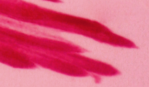
| This
character has arisen independently many times
in goby evolution and thus these genera are
not necessarily related. The state of the
pelvic fins is useful in larval identifications,
although it is quite clear that this character
can be inconsistent in larval stages and can
change at transition. The photograph at right
shows the divided pelvic fins in a larval
Psilotris
amblyrhynchus. |
|
| |
|
In some gobies, the divided pelvic fins are
clearly acquired after the larval phase. For
example, Coryphopterus
personatus larvae have fused pelvic
fins despite the fact that juveniles and adults
have separated pelvic fins. A closeup photograph
of the pelvic fin of a 7.6 mm SL larval C.
personatus at right clearly shows
the connecting membrane. Series of transforming
larvae show variable states of fusion of the
pelvic fins. It should be noted, however,
that the majority of larvae in the collections
have frayed fins and the state of fusion cannot
be evaluated. This is especially the case
for the difficult genus
Coryphopterus, where the pelvic-fin
morphology is, unfortunately, an important
species-level character. |
 |
 |
 |
Other
larval gobies, such as Gobulus
myersi and Psilotris
amblyrhynchus, can have partially-fused
pelvic fins. G.
myersi is an interesting contrast
to larval C.
personatus in that it shows the opposite
sequence of pelvic fin morphological changes:
it starts as a partially-divided fin in larvae
(left) and subsequently fuses in adults. |
|
| |
| The
only six-dorsal-spined species with divided pelvic
fins are a sub-group of Coryphopterus
(C. alloides,
C.
lipernes, C.
personatus, and C.
hyalinus). It is likely that the pelvic
fins of all of these species are not divided in
pre-transitional larvae. |
| |
| The
seven-dorsal-spined group with divided pelvic fins
is quite heterogeneous (with some rare and obscure
deep-water taxa), comprising Psilotris,
Varicus, Chriolepis,
and the individual species Pycnomma
roosevelti and very deep Robinsichthys
arrowsmithensis. |
| |
| Three
Caribbean goby species have partially-divided pelvic
fins (all seven-dorsal-spined): Gobulus
myersi, Psilotris
amblyrhynchus, and Gobiosoma
grosvenori. The latter is a member of the large
genus Gobiosoma
with otherwise fused pelvic fins and thus it is
unclear whether the larvae should be expected to
show any division in the pelvic fins. Gobulus
myersi adults have fused pelvic fins without
a frenum, but larvae clearly fitting this species
have partially-divided pelvic fins (D-VII,11-12
A-10-11). Two of the three other Gobulus
species have partially-divided pelvic fins
as adults (all in the eastern Pacific), and thus
the fused pelvic fin in adult G.
myersi may be a derived character. In contrast,
adult Psilotris
have divided pelvic fins and the presence of partially-fused
pelvic fins in larvae of Psilotris amblyrhynchus
may indicate that divided pelvic fins are a derived
character in that genus. |
| |
| In the
genus Psilotris, P.
batrachodes has the fewest fin rays with
modal D-9 A-7 Pect 16; P.
alepis has D-9-10-11 A-8-9
Pect 15, P. celsa
(originally "Psilotris celsus")
has D-9-11 A-9-10-11 Pect 16-17-19,
P.
boehlkei has D-10-11 A-10 Pect 16-18,
and P. kaufmani
has D-11 A-10-11 Pect 16-18-19. P.
amblyrhynchus has D-11-12 A-10-11
Pect 17-19. Psilotris
are scaleless. |
| |
| Pycnomma
roosevelti has a similar general appearance
and a modal fin-ray count of D-10 A-9 Pect 16 (and
later develops scales). (Gobiosoma
grosvenori also has modal D-10 A-9 (and Pect 17)
but is from a fused-fin genus and has only partially-divided
pelvic fins and a small pelvic frenum and a very
different body shape.) |
| |
| Chriolepis
and Varicus are
rare, obscure, and mostly deep-water gobies that
typically have divided and long pelvic fins and
large eyes. The species comprise Chriolepis
fisheri (the only relatively shallow water
species (can be found in sand tilefish mounds);
D-11-12 A-10-11 Pect 17-18, with two large spiny
basicaudal scales), Chriolepis
benthonis (over 150m, Gulf of Mexico, D-9
A-8 Pect 16), Chriolepis
bilix (described in 2013, over 60m, widespread,
D-12 A-11-12 Pect 19-20), and Chriolepis
vespa (deep, Gulf of Mexico, D-10 A-7-9 Pect
15-17). The related genus Varicus differs
by having unbranched pelvic-fin rays and comprises
Varicus bucca
(very deep-water, D-9-10 A-8 Pect 16-19), Varicus
marilynae (deep-water, Florida, D-9 A-8 Pect
16-18), and V. imswe
(deep-water, Belize, with pelvic fins extending
beyond the anal-fin origin and D-8 A-8 Pect 14-15).
A profoundly deep-water goby, Robinsichthys
arrowsmithensis, has D-VII,11 A-11 and is
distinctive with 22-23 pectoral-fin rays. |
| |
|
Note: fin-ray counts for the second dorsal fin and
the anal fin are total elements (spines plus rays)
and species are listed in rough order of increasing
anal-fin rays. |
|
|
|
|
|
|
|
|
| Diagnosis:
Clearly separated pelvic fins in the larval stage
and modal fin-ray counts of D-VII,9 A-7 (occ. 8)
and Pect 16 (occ.15), with usually two fewer anal-fin
rays than second-dorsal-fin rays, indicate Psilotris
batrachodes. This is the only Caribbean goby
with a modal count of as few as seven anal-fin elements.
(U) G19 |
|
| Analogues:
(post-anal fin solitary melanophore, small: <7
mm SL) |
|
| Description:
Body somewhat thick, long, and narrow with
a small round eye and a terminal mouth. Head relatively
broad and flattened. Pectoral fins long, dorsal
and anal-fin bases short and placed well back on
the body, caudal peduncle relatively short and narrowing.
Internal melanophores around the sacculus and on
the dorsal surface of the swim bladder as well as
around the gut near the vent. There is a large melanophore
on the ventral midline just after the last anal-fin
ray. Many individuals have a smaller matching melanophore
on the dorsal midline just after the last dorsal-fin
ray. Many individuals have a bubblewrap-like appearance
to the skin. |
|
|
|
| Psilotris
batrachodes larva |
| 6.0 mm SL |
| San Blas, Panama, SB84-522 |
| |
|
 |
| |
 |
| Psilotris
batrachodes adult |
| 13.3 mm SL |
| Saba Bank, Lesser Antilles |
| Photo by JT Williams |
|
 |
|
|
|
|
|
|
|
|
|
|
| Diagnosis:
A modal fin-ray count of D-VII,10 A-9 and
Pect 14-15 and divided pelvic fins indicates Psilotris
alepis (typically one fewer anal-fin ray than
second-dorsal-fin rays). The only Psilotris species
with fewer than 16 pectoral-fin rays are P. batrachodes and P.
alepis. P. batrachodes larvae have
fewer median-fin rays. Other gobies with divided
pelvic fins and matching median-fin ray counts have
more pectoral-fin rays and a different general appearance
(Pycnomma roosevelti and the deep-water
Chriolepis and Varicus).
(PE) G19b |
|
| Analogues:
(post-anal fin single melanophore, small: <7
mm SL) Among the taxa with divided pelvic fins
and the single melanophore behind the anal fin,
several types have small larvae. Larval Psilotris
alepis are separated from larval Pycnomma roosevelti by having
no dorsal melanophores, and from larval Gobulus myersi by having well-separated
and longer pelvic fins, a long pectoral fin reaching
almost to the vent, a wider caudal peduncle and
no abdominal midline melanophores. Larvae of Psilotris
batrachodes are similar and can be distinguished
by the lower fin-ray counts (especially the short
anal fin) and a narrower caudal peduncle. |
|
| Description:
Body long, narrow and somewhat thick, with a medium-sized
eye and a terminal mouth. Pectoral fins long, extending
to vent. Pelvic fins divided and short, extending
less than halfway to the vent. Dorsal and anal-fin
bases relatively short, caudal peduncle short and
wide. The caudal fin is wide and rounded. Lightly
marked along the lower body: melanophores along
the ventral midline at the pelvic-fin insertion
and a large, often dendritic, melanophore on the
ventral midline of the caudal peduncle just behind
the last anal-fin ray. Internal melanophores occur
at the dorsal surface of the swim bladder and around
the gut near the vent. Many individuals have a bubblewrap-like
appearance to the skin. |
|
|
|
| Psilotris
alepis larva |
| 6.3 mm SL |
| San Blas, Panama, SB86-625 |
| |
|
 |
| |
 |
| |
 |
| |
 |
| |
 |
| |
 |
|
|
|
|
|
|
|
|
|
| Diagnosis:
A modal fin-ray count of D-VII,10-11 (usually
11) A-10 Pect 16-18 and divided pelvic fins indicates
Psilotris boehlkei or P. celsa or
Chriolepis fisheri (typically
one fewer anal-fin ray than second-dorsal-fin rays).
Psilotris kaufmani can overlap the counts,
but usually has 11 anal-fin elements. C. fisheri can be distinguished
by having two basicaudal scales. Unfortunately the
11/10 median fin ray count is very common among
gobies and the state of the pelvic fins is therefore
a critical feature for diagnosis, but often difficult
to observe. Numerous Gobiosoma
and Tigrigobius
gobies overlap the counts, but have fused pelvic
fins- their overall morphology is often slightly
different, i.e. less likely to have a wide caudal
peduncle and rounded caudal fin and have a less
flattened head, but it is subtle. Gobies with six
dorsal spines and fused pelvics can share the fin-ray
count (and can have very similar adult markings),
including the sand gobies of Coryphopterus,
however they usually have equal numbers of dorsal
and anal fin rays and characteristically have relatively
narrow caudal penduncles and do not have the obvious
rounded caudal fin. |
|
| Description:
Larvae not identified. |
|
|
|
| Psilotris
boehlkei adult |
| 26.5 mm SL |
| Saba Bank, Lesser Antilles |
| Photo by JT Williams |
|
 |
|
|
|
|
|
|
|
|
| Diagnosis:
Mostly-divided pelvic fins and the modal
fin-ray count of D-VII,12 A-11 Pect 17-19 indicates
Psilotris amblyrhynchus and overlaps Gobulus myersi and Chriolepis fisheri (the latter
has fully divided pelvic fins and prominent modified
basicaudal scales). This larval type matches the
species description for P. amblyrhynchus. |
|
| Analogues:
(post-anal fin single melanophore, large:
>9 mm SL) Within the diverse solitary post-anal
fin melanophore group, there are few taxa with large
larvae: the other large larva is that of Nes longus. The similarity
is especially notable when larval Nes longus are missing
their anterior anal-fin base melanophores as well
as their caudal-fin base melanophores, which is
not uncommon. In that case, P. amblyrhynchus
larvae can be separated only by pelvic-fin morphology
(mostly divided pelvic fins with no frenum vs. fused
and an obvious frenum in Nes longus) and fewer
median-fin rays (usually 12/11 with a range of 11-12/9-11
vs. 12-14/11-13). Other members of the group are
much smaller: Psilotris, Gobulus myersi, and Pycnomma roosevelti with divided
pelvic fins and typically other distinctive melanophores,
and, with fused pelvic fins, some Gobiosoma and
Elacatinus,
along with Evermannichthys
(the latter also have a sharply-pointed
snout and spiny caudal peduncle scales). |
|
| Description:
Body thick with a large head that is flattened dorsally
and a large eye and terminal large mouth. Pectoral
fins medium length, not reaching the level of the
vent, pelvic fins medium length as well and mostly
separated, united near the base with a short membrane,
and no pelvic frenum. Dorsal and anal-fin bases
medium length, caudal peduncle relatively long and
somewhat narrow. Lightly marked along the lower
body: surface melanophores limited to a large prominent
dendritic melanophore at the ventral midline after
the last anal-fin ray. Internal melanophores are
present along the dorsal surface of the swim bladder.
Many individuals have a bubblewrap-like appearance
to the skin. |
|
|
|
| Psilotris
amblyrhynchus larva |
| 12.1 mm SL |
| Banco Chinchorro, Mexico,
coll. D. Jones |
| |
|
 |
| |
 |
| |
 |
| |
 |
|
|
|
|
|
|
|
|
| Diagnosis:
Modal fin-ray counts of D-VII,10 A-9 and Pect 16
and divided pelvic fins indicates Pycnomma roosevelti.
The appearance and fin-ray counts are quite similar
to the Psilotris, but a strong mode at this
count of fin elements excludes the Psilotris
species. P.
alepis larvae have fewer pectoral-fin rays
and lack the dorsal melanophore. Psilotris
boehlki and P. celsa both have a
strong modal anal-fin-ray count of 10 and are therefore
excluded. An adult Pycnomma roosevelti from
the Saba Bank matches the transitional larva in
morphology, especially the long body with a small
flattened head (and relatively small round eyes),
the placement of the anal fin far back on the body,
the wide caudal peduncle, and the rounded caudal
fin. The hunched-over appearance, with the head
placement mostly below the lateral midline of the
body, occurs in both the larvae and adult from Saba.
In addition, the larval melanophores on the body
at the end of the dorsal and anal fins match markings
at the same location on the Saba specimen. (PE)
G19a |
|
| Analogues:
(post-anal fin single melanophore, small:
<7 mm SL) Among the taxa with divided pelvic
fins and the single melanophore behind the anal
fin, several types have small larvae. Larvae of
Psilotris
batrachodes are most similar and often
have a post-dorsal fin spot as well (although it
is typically smaller than the ventral spot). When
the dorsal melanophore is present, Psilotris
batrachodes can be distinguished by the
lower fin-ray counts (especially the short anal
fin) and a narrower caudal peduncle. Psilotris
alepis larvae have only a ventral spot and
Gobulus
myersi have abdominal ventral midline spots. |
|
| Description:
Body somewhat thick, long, and narrow with
a relatively small round eye and a terminal mouth.
Larvae are usually hunched-over, with the head mostly
bent below the level of the lateral midline of the
body. Head relatively broad and flattened, not much
wider than the caudal peduncle. Pectoral fins long,
dorsal and anal-fin bases short and placed well
back on the body, caudal peduncle relatively wide.
Internal melanophores around the sacculus and on
the dorsal surface of the swim bladder. A surface
melanophore around the gut near the vent and large
solitary melanophores on the caudal peduncle placed
just behind the last dorsal and anal-fin rays. Transitional
larvae become more flattened and develop tiny melanophores
on the mid-upper and lower jaw and a scattering
of leukophores on top of the head between the eyes. |
|
|
|
| Pycnomma
roosevelti larva |
| 6.0 mm SL |
| Yucatan, Mexico, 240306 |
| coll. by Lourdes Vasquez
et al. |
|
 |
| Pycnomma
roosevelti larvae |
| 6.0 mm SL |
| Yucatan, Mexico, 240306 |
| coll. by Lourdes Vasquez
et al. |
|
 |
| |
|
| Pycnomma
roosevelti larva |
| 6.0 mm SL |
| divided pelvic fins |
| Yucatan, Mexico, 240306 |
| coll. by Lourdes Vasquez
et al. |
|
 |
| Pycnomma
roosevelti transitional larva |
| 5.5 mm SL |
| San Blas, Panama, SB84-624a |
| |
|
 |
| Pycnomma
roosevelti adult |
| 14.6 mm SL |
| Saba Bank, Lesser Antilles |
| Photo by JT Williams |
|
 |
| |
|
| |
|
|
|
|
|
|
|
|
|
| Diagnosis:
Modal fin-ray counts of D-VII,11 A-10 and
Pect 15 and partially-divided pelvic fins indicate
Gobulus myersi and barely overlaps the range
of some Psilotris
species These genera have one fewer anal-fin
ray (sometimes two) than second-dorsal-fin rays.
Pelvic fins can be fused or halfway-fused in these
larvae. The short pectoral fins and pelvic fins
without a frenum, rounded caudal fin, broad head
and stocky body of this larval type fits with Gobulus
myersi (or Psilotris)
rather than the other gobies with these fin-ray
counts and fully-fused pelvic fins (i.e. some Gobiosoma and
Elacatinus).
The fin-ray count of this larval type borders the
range but does not match Psilotris
(too high for P. alepis; modal pectoral-fin rays
too low for P. boehlkei and P. celsa).
Confirming the identification as G. myersi
is the white spotting on the upper half of the body
from the top of the head to the caudal fin in transitional
larvae: this species is unusual in being bicolored
with light above and dark below (the common name
is paleback goby). Gobulus is a genus with
only four species; this one representative in the
Atlantic and three endemic to the eastern Pacific
region. Although this Atlantic species is reported
to have fused pelvic fins (without a frenum) as
adults, two of the Pacific Gobulus species
have partially-fused pelvic fins (without a frenum),
as do these larvae. fin-ray counts in this larval
type often vary from the mode: second-dorsal-fin
elements are often 10 or 12, anal-fin elements are
often 9 or 11 and pectoral-fin-ray counts are often
14 or 16. (Chriolepis
can share the median-fin ray count but
should have more pectoral-fin rays, longer and separate
pelvic fins, and a larger eye). (PE) G8a |
|
| Analogues:
(post-anal fin solitary melanophore, small:
<7 mm SL) |
|
| Description:
Body shape ranges from thin, long and narrow in
earlier-stage larvae to thicker with a large head,
medium-sized eye and a terminal large wide mouth.
Pectoral fins short, reaching about halfway to the
vent. Pelvic fins without a pelvic frenum and can
be fused or halfway fused, and short, extending
clearly less than halfway to the vent, 4-5 procurrent
caudal-fin rays. Dorsal and anal-fin bases relatively
short, caudal peduncle short and narrowing. The
caudal fin is short, wide and rounded. Lightly marked
along the lower body: melanophores along the ventral
midline sometimes at the isthmus, then just forward
of the pelvic-fin insertion, followed by from one
to seven additional melanophores along the abdominal
midline always ending with a melanophore just forward
of the vent. There are no melanophores along the
anal-fin base. There is a prominent large, often
dendritic, melanophore on the ventral caudal peduncle
midline just behind the last anal-fin ray. Melanophores
occur internally along the dorsal surface of the
swim bladder and around the gut near the vent (no
melanophores at the sacculus). Series of transitional
larvae show development of the eye from a somewhat
narrowed vertical oval to round (in most specimens
the eye is round). Transitional larvae develop a
dark stripe through the eye back to the operculum,
melanophores on the lower jaw, scattered around
and within the thorax, and in a stripe along the
sides of the abdominal wall to the vent and continuing
along the base of the anal fin and caudal peduncle
to curve around the base of the lower and central
caudal-fin rays. There is a conspicuous speckling
of leukophores (and an absence of any melanophores)
over the top of the head and extending along the
dorsum of the body onto the caudal fin. Some individuals
have a bubblewrap-like appearance to the skin. |
|
|
|
| Gobulus
myersi larva |
| 5.4 mm SL |
| San Blas, Panama, SB86-407 |
| |
|
 |
| |
 |
| |
 |
| Gobulus
myersi larva |
| 5.5 mm
SL |
| seven midline abdominal
melanophores |
| San Blas, Panama,
SB86-1103 |
| |
|
 |
| |
 |
| Gobulus
myersi larva |
| 5.7 mm SL |
| slightly narrowed vertical
eye |
| San Blas, Panama,
SB87-219 |
| |
|
 |
| Gobulus
myersi larvae |
| 5.2 and 5.5 mm SL |
| San Blas, Panama, SB86-503 |
| |
|
 |
| Gobulus
myersi larva |
| 5.5 mm SL |
halfway fused pelvic
fins
and short pectoral fins |
| San Blas, Panama, SB86-503 |
| |
|
|
| |
 |
| Gobulus
myersi larva |
| 5.8 mm SL |
| D-VII,12 A-11 Pect 15 |
| San Blas, Panama, SB86-503 |
| |
|
 |
| Gobulus
myersi transitional larva |
| 6.0 mm SL |
| San Blas, Panama, SB86-501 |
| |
|
 |
| |
 |
| |
 |
| Gobulus
myersi transitional larva |
| 6.0 mm SL |
| leukophores on dorsal
side |
| San Blas, Panama, SB87-224 |
| |
|
 |
| Gobulus
myersi transitional larvae |
| 6.0 and earlier-stage,
4.9 mm SL |
| San Blas, Panama, SB87-224 |
| |
|
 |
|
|
|
|
|
|
|
|
| Diagnosis:
Modal fin-ray counts of D-VII,11-12 A-10-11 and
Pect 17-18 and divided pelvic fins indicates Chriolepis
fisheri. Only this species has two prominent
spiny basicaudal scales, and they are present on
late-stage larvae. |
|
| Analogues:
(post-anal fin single melanophore, small:
<7 mm SL) |
|
| Description:
|
|
|
|
|
|
|
|
| Chriolepis/Psilotris
sp. 23 (?) |
|
|
|
|
|
|
|
|
|
|
|
|
Diagnosis:
An individual larva with a robust body and large
eye and the fin-ray count of D-VII,10 A-10 and
Pect 16 falls within the range of a number of
regional species. The state of the pelvic fins
is uncertain but there is no indication of a frenum
and, with this fin-ray count, it is likely that
the pelvic fins in this larval type are divided,
since fused-pelvic-fin gobies rarely have as few
as 10 second-dorsal-fin elements: only Barbulifer
and Gobiosoma
grosvenori have 10, but both have the typical
one-fewer-anal than dorsal-fin elements. In addition,
Barbulifer
larvae are morphologically quite different and
larval Gobiosoma
are lightly-marked and smaller (note that rare
individuals of Gobiosoma spes and
G. yucatanum would also overlap the fin-ray
count of this larva, but neither would resemble
this larva).
Among the divided-pelvic-fin
species, a number have overlapping fin-ray counts
to this larval type, including Psilotris celsa,
P. boehlkei, P. kaufmani,
and Pycnomma roosevelti. Species of
Chriolepis have similar (but not the same)
fin-ray counts. Larvae of P. roosevelti are quite different
and have small eyes and a flattened head. Psilotris boehlkei do not appear
similar: the Saba adult is relatively lightly
marked with a pale body marked only by short midline
internal linear melanophores, melanophores in
a patch under the lower jaw, a large patch below
the eye, a patch on the upper pectoral-fin base
and stripes back from the upper eye (notably,
these markings are similar to the common Coryphopterus
sand gobies). On the Saba fish, there are two
stripes across the spinous dorsal fin, black edging
to the soft dorsal-fin membranes and around the
caudal fin and mostly dark membranes on the anal
fin. Notably there are no obvious ventral midline
melanophores along the anal fin and caudal peduncle
which are prominent on this larval type. Psilotris
kaufmani and Psilotris celsa cannot
be excluded as possible candidates, and the former
shares the large eye and bulky body of this larva.
Chriolepis spp. cannot be ruled out, but
the larva differs from larval Chriolepis fisheri
in appearance and fin-ray counts and lacks the
spiny basicaudal scales. (PE) G23
|
|
|
Analogues:
(heavy ventral markings) This larval type
shares the dendritic melanophores along the ventral
midline with the superficially-similar Bathygobius mystacium and B. soporator larval types.
This larval type differs in having the markings
continue onto the caudal peduncle and base of
the caudal fin, an area spared in transitional
Bathygobius larvae. In addition,
this larval type has seven first-dorsal-fin spines
vs. six in Bathygobius.
|
|
|
Description: Body
relatively thick, long and narrow with a large
eye and a terminal large mouth. Pectoral and pelvic
fin length uncertain, without an obvious pelvic
frenum. Dorsal and anal-fin bases medium-length
and caudal peduncle medium-length and relatively
wide, 6-7 procurrent caudal-fin rays (6 spindly).
Heavily marked; along the ventral midline there
are large deep internal melanophores underlying
the isthmus and forward of the pelvic-fin base.
The abdominal midline is unmarked. There is a
heavy continuous line of large dendritic melanophores
along the lateral abdominal wall continuing along
the base of the anal fin and ventral midline of
the caudal peduncle onto the caudal fin. Melanophores
line the bases of most of the caudal-fin rays,
extending out along the segmented rays. Large
dendritic melanophores lie along the dorsal midline
of the caudal peduncle and around the base of
the soft dorsal fin near the fourth element and
the last two elements. The lateral abdominal wall
row merges internally with pigment overlying the
swim bladder and extends forward into the head.
Head markings include melanophores directly above
the eyeball, near the tip of the upper jaw, between
the eye and the mid-upper jaw, and directly below
the eye and just forward of the angle of the jaw,
which also has the typical associated melanophore.
The lower jaw has a complex arrangement of melanophores:
there are a pair outlining the lower edge of the
dentary on each side of the midline, a large dendritic
pair on each side below the dentary and then one
at the ventral midline halfway down the lower
jaw and a pair just forward of the pair at the
angle of the jaw. There are scattered melanophores
overlying the iris. The eye is large and round.
|
|
|
|
| Chriolepis/Psilotris
sp. larva |
| 7.4 mm SL |
| San Blas, Panama, SB81-196 |
| |
|
 |
| |
 |
| |
 |
| |
 |
|
|
|
|
|
| |
|
|
|
|
|
| |
|
 |
|
All contents © copyright 2006-2014
All rights reserved
www.coralreeffish.com by
Benjamin Victor
|
|
|
|
|
|