The
dartfishes of the family Ptereleotridae have been
taxonomically mobile in recent years and some taxonomists
now include them in the wormfish family
Microdesmidae. I place them here alongside the
family Eleotridae because they are also gobioids
and share the clearly-divided pelvic fins of the
eleotrids. Larval
ptereleotrids most closely resemble the "long"
larvae of my Group
4 gobies. There are only two dartfishes in the
region, a pair of sibling species that vary only
slightly in color: their larvae are likely identical.
The sleepers of
the family Eleotridae (some use Eleotrididae) are
similar to gobies but have divided and well-separated
pelvic fins (photograph of the separated pelvic
fins in
a
12.8 mm SL larval Eleotris
amblyopsis). There are six Caribbean
genera, most are probably monospecific. The
eleotrid species are widespread in the Caribbean,
except for Leptophilypnus
fluviatilis, an estuarine species
recorded only from the central American coastline
from Honduras to Panama and another obscure
species, Leptophilypnus guatemalensis
native to far inland freshwater rivers in
Guatemala.
Eleotrids are typically found in tropical
freshwater habitats, but they do penetrate
brackish and mangrove environments. Some species
get large as adults and can even become gamefishes
in the major river systems of central America.
Their larvae, however, are small to medium-sized
and exhibit similar body shapes and marking
patterns to the larvae of their goby relatives.
Larval
eleotrids share many basic characters of larval
gobies. While most of the true gobies have
fused pelvic fins, several genera have divided
pelvic fins like the eleotrids (although they
do not have the fins completely separated
at the base as do the sleepers). pelvic-fin
morphology is not always easily apparent on
small larvae, but fortunately there are only
a few species of eleotrids in the Caribbean
and larval eleotrids do have a somewhat different
appearance from the usual goby gestalt.
Most
eleotrid larvae share a distinctive suite
of characters. They have long ventral midline
streaks of melanophores that extend onto the
abdomen. Most also have linear internal melanophores
extending up from the anal-fin base along
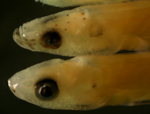
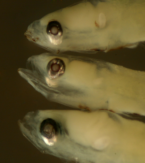
myomere edges. Pre-transitional stages usually
have odd-shaped narrowed eyeballs, some with
unusual pigmented membranes over the iris
that can expand to essentially cover the shiny
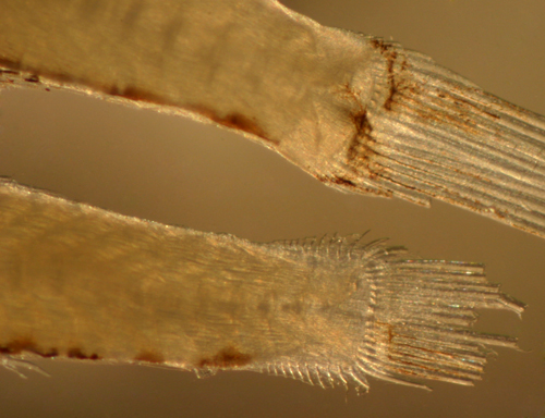
surface completely. Also unusual is the high
number of procurrent caudal-fin rays, up to
14, in several eleotrid species; true gobies
almost always have 10 or fewer, often many
fewer. The goby exceptions are the larvae
of the river gobies of Sicydium
and Awaous.
Interestingly, the common factor is freshwater
habitat; the high number of procurrent caudal-fin
rays is likely an adaptation for living in
fast-flowing streams. Eleotrid larvae also
exhibit some of the more dramatic eye-shape
changes during development and at transition
found in larval fishes.
Eleotrid
larvae tend to share basic melanophore patterns
and general morphology, and fin-ray count
differences are slight. There is also a marked
degree of variation within species, making
species identifications more difficult. Some
characters common in one larval type will
occur occasionally (or later in transition)
in another larval type; for example, the characteristic
melanophore patterns along the jaws of Eleotris
amblyopsis larvae match closely those
found on the late transitional larvae of Erotelis
smaragdus. Typically, a suite of characters
in combination serve to distinguish the larval
types and unite transitional series.
The
literature reports of fin-ray counts of sleepers
can differ by two fin rays or more, and sometimes
disagree on whether there are equal numbers
of dorsal and anal-fin rays or more or less.
The literature on larval sleepers is also
not helpful, since features common to the
entire family are typically cited as unique
to one species or other and line drawings
omit other diagnostic features (likely a result
of inadequate sample sizes of highly-variable
larvae).
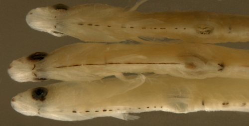
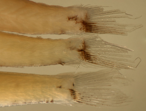
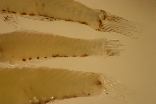
Complex
neuromast patterns develop on the head, body
and caudal fins of late transitional eleotrid
larvae. These patterns are more developed
on juveniles and adults and are commonly used
for taxonomic studies. Unfortunately, the
neuromasts can be hard to highlight on most
transitional larvae (photograph below, from
the top, larval Erotelis
smaragdus, Gobiomorus
dormitor, and Eleotris
amblyopsis).
Dormitator maculatus
Diagnosis:
Modal fin-ray counts of D-VII,9 A-10 Pect-14
indicate Dormitator
maculatus. This is the only Caribbean eleotrid
with more anal-fin elements than second-dorsal-fin
elements (the eastern Pacific sleeper, Gobiomorus
maculatus, shares this feature with a modal
fin-ray count of D-VI,10 A-11). D.
maculatus is also distinguished from the
other eleotrids by the low pectoral-fin-ray count
and the relatively low numbers of procurrent caudal-fin
rays (7-9 vs. 10 or more in several other eleotrids).
D. cubanus has
been described as the Cuban form and D.
lophocephalus as the Surinamese form, although
the validity of these species is uncertain. (U)
G2
Analogues:
Larval Dormitator maculatus
share a long thin body with relatively short dorsal
and anal-fin bases and a long ventral midline streak
from the isthmus to the mid-abdomen with the other
eleotrid larvae. However, the larvae are distinctive
in having relatively few procurrent caudal-fin rays.
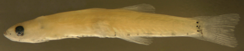
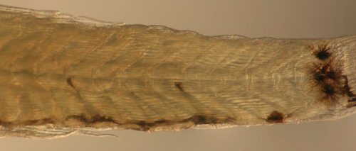
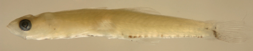
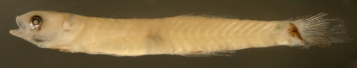
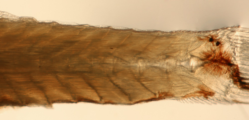
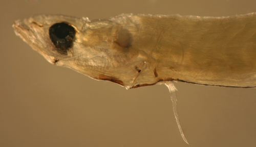
Description:
Body thin, long and narrow with a relatively small,
often oval, eye and a terminal medium-sized mouth.
Pelvic fins separate and short, pectoral fins short,
dorsal and anal-fin bases short, caudal peduncle
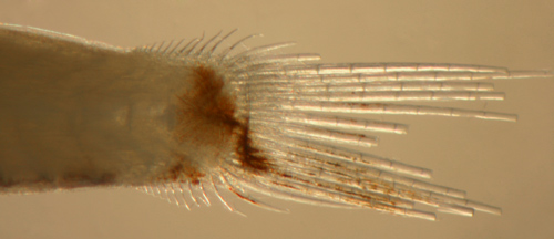
long and sharply narrowing, procurrent caudal-fin
rays 7-9. Markings mostly along the ventral midline:
melanophores usually as streaks extending from the
isthmus to the mid-abdomen, ending at the swim bladder
(which is the full thickness of the abdomen and
provides a clear view of the retroperitoneum); there
is often an additional small melanophore on the
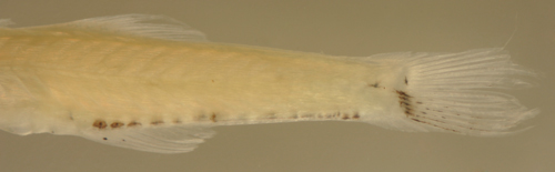
promontory just forward of the vent. There are four
to six large melanophores along the bases of the
last eight anal-fin rays (paired) and continuing
as a streak of seven or eight large melanophores
along the ventral peduncle ending at the start of
the lower procurrent caudal-fin rays (the last in
the series often larger and extends deeper into
the musculature). There are melanophores at the
base of most of the lower segmented caudal-fin rays,
some variably extending along the length of the
lower segmented caudal rays as well as sometimes
on the distal half of the central segmented caudal-fin
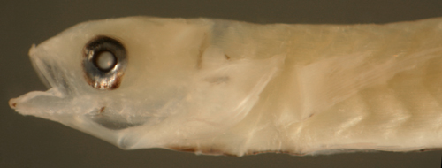
rays. Head markings consist of melanophores outlining
the lower rim of the tip of the dentary of the lower
jaw. Varying proportions of individuals in different
daily collections possess a melanophore at the angle
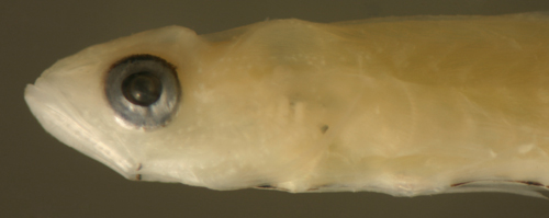
of the jaw, from none to most. There is usually
a prominent "eyebrow" membrane lined with melanophores
overlying the dorsal aspect of the upper third of
the eyeball. Additional melanophores occur on the
occasional individual in any combination of the
following: on the proximal membranes of the anal-fin
elements (usually the third and fourth, rarely all),
overlying the cleithrum visible upon lifting the
operculum, or on the outer operculum near the lower
edge on each side. Internal melanophores typically
surround the saccule (in most well-developed larvae)
and are present along the dorsal surface of the
swim bladder (none around the gut near the vent).
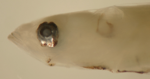
Series of transitional larvae show development of
the eye from a narrowed vertical oval, sometimes
tilted slightly forward (occasionally backward),
with the pupil off-center dorsally, a small posterior-inferior
extension of the iris, and often a dorsal indentation
in the iris) to almost rounded, sometimes tilted
sharply backward.
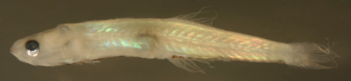
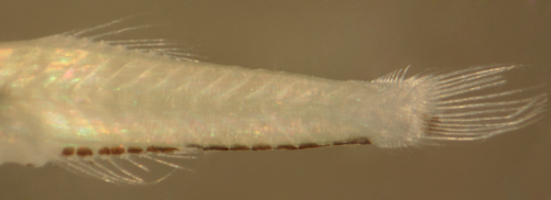
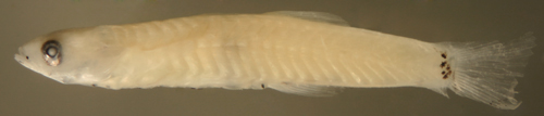
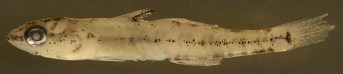
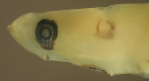
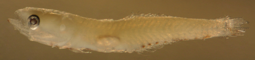
Dormitator
maculatus larva
7.9 mm SL
San Blas, Panama, SB86-1001
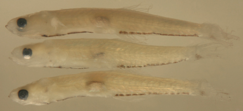
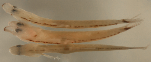
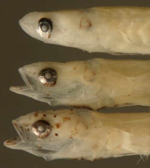
Dormitator
maculatus larvae
7.9, 7.6, and 8.0 mm
SL
note eye shape variation
San Blas, Panama, SB86-1001
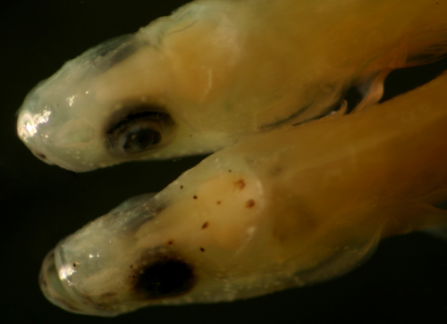
Dormitator
maculatus larvae
7.9 and 8.0 mm SL
variation in markings
including
opercular (above), cleithral (below),
abdominal promontory and
anal-fin ray melanophores
San Blas, Panama, SB86-1002
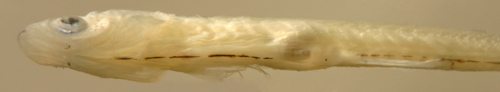
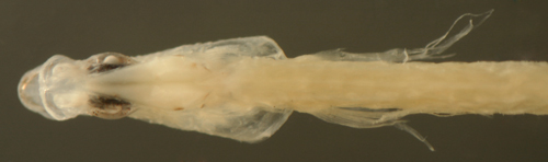
Dormitator
maculatus larva
7.2 mm SL
note stubs of separate
pelvic fins
San Blas, Panama, SB86-927
Dormitator
maculatus larva
8.1 mm SL
prominent "eyebrow"
membrane
San Blas, Panama, SB86-1001
Dormitator
maculatus larva
8.2 mm SL
eye tilted backwards
San Blas, Panama, SB86-1030
Dormitator
maculatus larva
8.1 mm SL
San Blas, Panama, SB86-1030
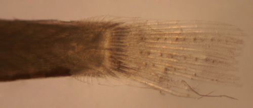
Dormitator
maculatus larva
7.4 mm SL
note three rows of
caudal-fin papillae
San Blas, Panama, SB86-1006
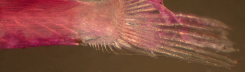
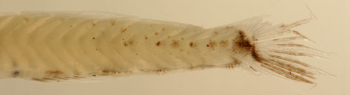
Dormitator
maculatus larva
8.1 mm SL
relatively few procurrent
caudal-fin rays
San Blas, Panama, SB86-1001
Eleotris amblyopsis
Diagnosis:
Modal fin-ray counts of D-VI,9 A-9 Pect-16
indicate Eleotris amblyopsis.
E. perniger is almost identical (and can
co-occur with E. amblyopsis)
but is reported to have a mode of 18 pectoral-fin
rays with a range from 16 to 19 vs. a mode of 16
and a range of 15-18 for E.
amblyopsis (Pezold 2002). The larval type
described here has mostly 15 and 16 pectoral-fin
rays. This is the only eleotrid genus with as few
as 8 or 9 second dorsal and anal-fin elements and
equal numbers. This taxon historically has been
variably split: although E.
pisonis has been considered the widespread
Caribbean species and books refer to it as such,
recent work by Pezold (2002) indicates that the
true E. pisonis
is South American, ranging from Orinoco-Venezuela
to Brazil. Instead, E.
amblyopsis occurs along the continental margin
from the US to Venezuela and E.
perniger is mostly of the Antilles, but co-occurs
with E. amblyopsis
along the Central American coast. Since the species
are morphologically virtually identical, the larvae
may be indistinguishable without DNA sequencing.
Two larvae caught together on one day are larger
than virtually all of the others, about 14.5 mm
SL, and both have 17 pectoral-fin rays; it is possible
that they represent E.
perniger, although DNA sequence analysis
will be required for confirmation. (U) G4
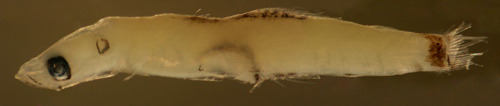
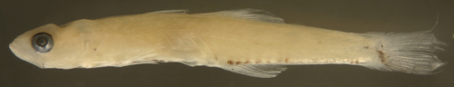
Analogues:
The eleotrid larvae share a long thin body with
relatively short dorsal and anal-fin bases, numerous
procurrent caudal-fin rays, and a long ventral midline
streak from the isthmus to the mid-abdomen. Larval
Eleotris amblyopsis
are distinguished by a wide caudal peduncle, a pigmented
end to the caudal peduncle, and short wing-like
dorsal and anal fins with no melanophores on the
membranes.
Description:
Body somewhat thick, long and narrow (pretransitional
individuals can be relatively wide for this family)
with a medium eye and a terminal medium-sized mouth.
Pectoral fins short, reaching only about halfway
to vent. Pelvic fins short and clearly separate.
Dorsal and anal-fin bases very short and the front
and middle rays are longer than the back rays making
a triangular outline to the fin, caudal peduncle
relatively wide and long and there are many procurrent
caudal-fin rays in a distinct fleshy fold, from
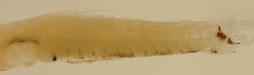
12-14 in this larval type. Markings mostly along
the ventral midline: melanophores usually as streaks
(but often contracted and appear as spots) extending
from the isthmus most of the way to the vent (if
spots, there are 10 to 15), but absent below the
swim bladder (which is the full thickness of the
abdomen and provides a clear view of the retroperitoneum).
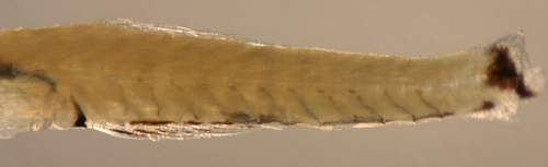
There is an irregular paired row of melanophores
along the bases of the last seven or eight anal-fin
rays continuing as three or four streaks along the
ventral midline of the peduncle ending at the start
of the lower procurrent caudal-fin rays. The end
of the caudal peduncle is uniformly covered by a
wide bar of large melanophores, usually not extending
all the way to the lower procurrent caudal rays
or to the edge of the upper procurrent caudal-fin
rays. Melanophores extend out along the proximal
lower segmented caudal-fin rays and rarely along
the distal portions of the central or upper segmented
caudal rays. Only a rare individual has a small
melanophore on the dorsal midline of the caudal
peduncle forward of the start of the procurrent
caudal-fin rays. Head markings on all individuals
include melanophores at the angle of the jaw, along
the dentary at the tip and below the dentary just
to the side of the tip of the lower jaw. Head markings
on most individuals include melanophores
on the rim of the mid-maxilla, on the dentary of
the mid-lower jaw, and below the nasal bones. Prominent
melanophores extend over the surface of the iris
on the dorsal half of the eyeball and paired around
the base of the eyeball and, when expanded, melanophores
almost cover the iris. Occasional individuals have
a hidden melanophore overlying the cleithrum visible
upon lifting the operculum (rarely paired). Internal
melanophores surround the saccule and are present
along the dorsal surface of the swim bladder and
paired around the gut near the vent. There is a
linear internal melanophore above the pelvic girdle,
perhaps along the ventral postcleithrum and a few
internal melanophores are often present around vertebral
bodies near the tail and sometimes continuing along
the hemal spine down to meet the midline melanophores.
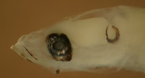
Series of transitional larvae show the body narrowing
and thickening and development of the eye from a
moderately narrowed vertical oval, tilting forward,
with the pupil off-center dorsally and a slight
posterior-inferior extension of the iris (there
is also occasionally a marked dorsal and ventral
indentation of the iris) to fully round. Transitional
larvae develop a pattern of several discrete melanophores
on the top of the head, at the midline behind the
upper lip, and forward of the eye.
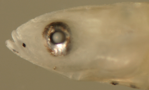
Eleotris
amblyopsis larva
12.0 mm SL
San Blas, Panama, SB86-1008
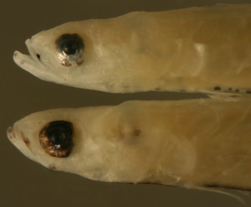
Eleotris
amblyopsis larva
12.3 mm SL
early-stage narrow
eye and wide body
San Blas, Panama, SB86-627
Eleotris
amblyopsis larva
13.3 mm SL
later rounder eye and
narrower body
San Blas, Panama, SB86-1001
Eleotris
amblyopsis larva
13.2 mm SL
round eye, no transitional
melanophores
San Blas, Panama, SB84-523
Eleotris
amblyopsis larva
12.3 mm SL
San Blas, Panama, SB86-627
Eleotris
amblyopsis larva
12.0 mm SL
short fin bases and
triangular fin shapes
San Blas, Panama, SB86-1008
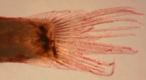
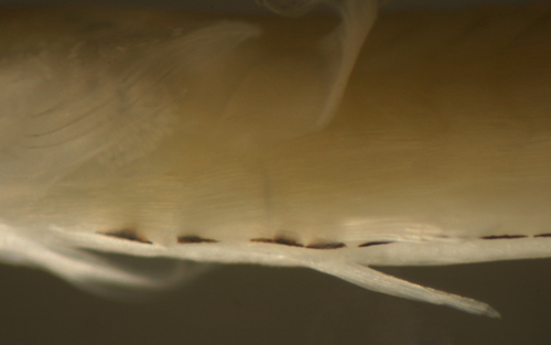
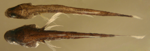
Eleotris
amblyopsis larva
12.2 mm SL
note 12-14 procurrent
caudal-fin rays,
melanophore at dorsal midline
San Blas, Panama, SB86-1002
Eleotris
amblyopsis larva
13.3 mm SL
three rows of sensory
papillae neuromasts
San Blas, Panama, SB86-1001
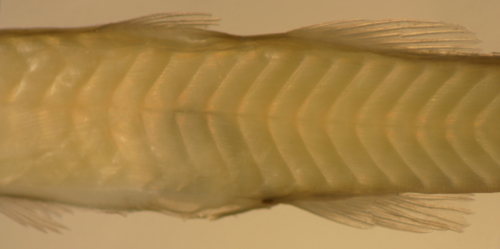
Eleotris
amblyopsis larva
11.7 mm SL
internal melanophores
along vertebral
bodies and hemal spines
San Blas, Panama, SB86-927
Eleotris
amblyopsis larva
12.3 mm SL
San Blas, Panama, SB86-627
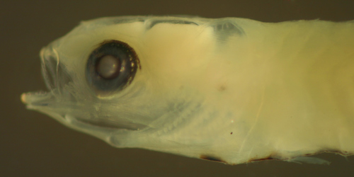
Eleotris
amblyopsis larva
12.4 mm SL
note indentations in
iris
San Blas, Panama, SB86-1006
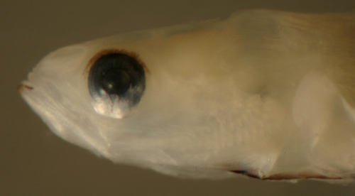
Eleotris
amblyopsis larva
13.3 mm SL
note round eye
San Blas, Panama, SB86-1030
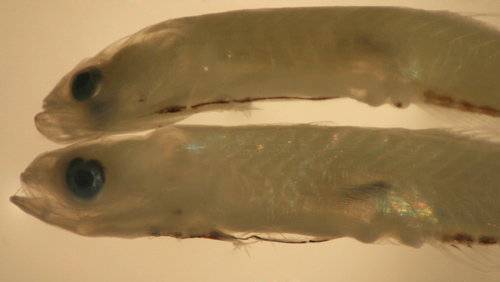
Eleotris
amblyopsis larvae
12.3 and 12.4 mm SL
note melanophores contracted
vs. expanded, and sensory papillae
San Blas, Panama, SB86-1001
Eleotris
amblyopsis larvae
12.3, 12.4, and 12.5
mm SL
melanophores contracted
vs. expanded
San Blas, Panama, SB86-1001
Eleotris
amblyopsis larva
and early transitional larva below
12.6 mm SL
San Blas, Panama, SB86-627
Eleotris
amblyopsis transitional larva
12.2 mm SL
note expanded melanophores
over the iris
San Blas, Panama, SB86-1002
Eleotris
amblyopsis transitional larva
13.2 mm SL
note neuromast development
on head,
and melanophore overlying cleithrum
San Blas, Panama, SB81-019
Eleotris
amblyopsis larva above
vs. Gobiomorus dormitor larva below
12.1 mm SL
note neuromast pattern
differences
San Blas, Panama, SB86-1002
Eleotris perniger?
Diagnosis:
Modal fin-ray counts of D-VI,9 A-9 Pect-18
indicate Eleotris perniger.
This species is almost identical (and can co-occur)
with E. amblyopsis,
but is reported to have a mode of 18 pectoral-fin
rays with a range from 16 to 19 vs. a mode of 16
and a range of 15-18 for E.
amblyopsis (Pezold 2002). This is the only
eleotrid genus with as few as 8 or 9 second dorsal
and anal-fin elements and equal numbers. Taxonomy
is unresolved in this genus; E.
perniger is mostly of the Antilles, but reportedly
co-occurs with E. amblyopsis
along the Central American coast. This larval type
is from off Yucatan and has distinctively different
markings along the dorsal-fin bases compared to
typical E. amblyopsis
larvae collected from Panama. It is identified as
E. perniger
pending DNA identification.
Analogues:
The eleotrid larvae share a long thin body with
relatively short dorsal and anal-fin bases, numerous
procurrent caudal-fin rays, and a long ventral midline
streak from the isthmus to the mid-abdomen.
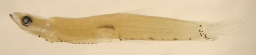
Description:
Body somewhat thick, long and narrow with a medium
eye and a terminal medium-sized mouth. Pectoral
fins short, reaching only about halfway to vent.
Pelvic fins short and separate. Dorsal and anal-fin
bases very short and the front and middle rays are
longer than the back rays making a triangular outline
to the fin, caudal peduncle relatively wide and
long and there are many procurrent caudal-fin rays
in a distinct fleshy fold, from 12-14 in this larval
type. Dorsal markings are prominent along the dorsal-fin
bases, as paired melanophore rows running alongside
the base of both fins. Along the ventral midline,
melanophores occur in streaks extending from the
isthmus most of the way to the vent, but absent
below the swim bladder (which is the full thickness
of the abdomen and provides a clear view of the
retroperitoneum). There is an irregular paired row
of melanophores along the bases of the last seven
or eight anal-fin rays and continuing as three or
four streaks along the ventral midline of the peduncle
ending at the start of the lower procurrent caudal-fin
rays. The end of the caudal peduncle is uniformly
covered by a wide bar of large melanophores, usually
not reaching the bases of the upper and lower procurrent
caudal rays. Melanophores extend out along the proximal
lower segmented caudal-fin rays. Head markings include
melanophores at the angle of the jaw, along and
beside the dentary of the lower jaw and outlining
the maxillary. Prominent melanophores extend over
the surface of the iris on the dorsal half of the
eyeball and paired around the base of the eyeball
and, when expanded, melanophores almost cover the
iris. Internal melanophores surround the saccule
and are present along the dorsal surface of the
swim bladder extending down to the vent. There is
a linear internal melanophore above the pelvic girdle,
perhaps along the ventral postcleithrum, and a few
internal melanophores are often present around vertebral
bodies near the tail and sometimes continuing along
the hemal spine down to meet the midline melanophores.
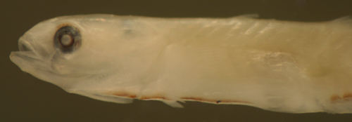
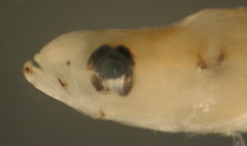
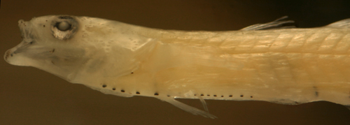
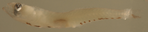
Eleotris
perniger? larva
8.1 mm SL
Yucatan, Mexico, 280306
coll. by Lourdes Vasquez
et al.
Gobiomorus dormitor
Diagnosis:
Modal fin-ray counts of D-VI,11 (or 10) A-10
Pect-16 or 17 indicate Gobiomorus
dormitor or Erotelis
smaragdus. These genera have overlapping
fin-ray counts and, given the inconsistent ranges
reported in the literature, these cannot be relied
on to distinguish the taxa. Leptophilypnus
fluviatilis, from Panama, overlaps the dorsal
and anal-fin-ray counts (with 10/10), but has 18-19
pectoral-fin rays. Larvae and recruits of Erotelis
smaragdus differ in having a small eye and
a row of melanophores along the base of the anal-fin
membranes. The larvae of the eastern Pacific sibling
species, Gobiomorus
maculatus, shares the melanophore pattern.
G4a
Analogues:
The eleotrid larvae share a long thin body with
relatively short dorsal and anal-fin bases, numerous
procurrent caudal-fin rays, and a long ventral midline
streak from the isthmus to the mid-abdomen. Larval
Gobiomorus dormitor
are distinguished by a particularly large eye (as
transition approaches), melanophores on the first
anal-fin membrane only, and the early appearance
of melanophores at the base of some of the upper
caudal-fin rays. The latter develop only during
transition in Eleotris.
Description:
Body somewhat thin, long and narrow with a large
eye and a terminal medium-sized mouth. Pectoral
fins short, reaching only about halfway to vent.
Pelvic fins short and clearly separate. Dorsal and
anal-fin bases very short and the front and middle
rays are longer than the back rays making a triangular
outline to the fin, caudal peduncle relatively narrow
and long and there are many procurrent caudal-fin
rays in a distinct fleshy fold, from 11-14 in this
larval type. Markings mostly along the ventral midline:
melanophores usually as streaks (but often contracted
and appear as spots) extending from the isthmus
most of the way to the vent (about 10), ending at
the swim bladder (which is the full thickness of
the abdomen and provides a clear view of the retroperitoneum),
most individuals have an additional midline melanophore
on the promontory just forward of the vent. There
is a row of five melanophores along the bases of
the last seven or eight anal-fin rays (variably
paired) and several more continuing mostly as a
streak along the ventral peduncle ending at the
start of the lower procurrent caudal-fin rays. There
are melanophores on the membranes only between the
first and third anal-fin elements. Melanophores
are concentrated at the base of the lower caudal-fin
segmented rays extending out along the length of
the rays. Unlike pre-transitional larvae of the
other regional larval eleotrids discussed, there
are melanophores at the base of soem or all of the
upper caudal-fin segmented rays as well. The caudal
peduncle has deep internal melanophores around the
point of flexion of the spine which can connect
to the caudal-fin base melanophores. Head markings
comprise melanophores at the angle of the jaw and
along the lower jaw at the tip and below the dentary
just to the side of the tip (no markings on the
upper jaw). There is often an "eyebrow" membrane
lined with melanophores overlying the dorsal aspect
of the upper third of the eyeball. There is a melanophore
overlying the cleithrum visible upon lifting the
operculum (paired). Internal melanophores surround
the saccule and are present along the dorsal surface
of the swim bladder and paired around the gut near
the vent. Some individuals show several of the ventral
midline melanophores extending internally along
the hemal spine in the body segments posterior to
the the vent. Series of transitional larvae show
development of the eye from a moderately narrowed
vertical oval with a slight posterior-inferior extension
of the iris to fully round.
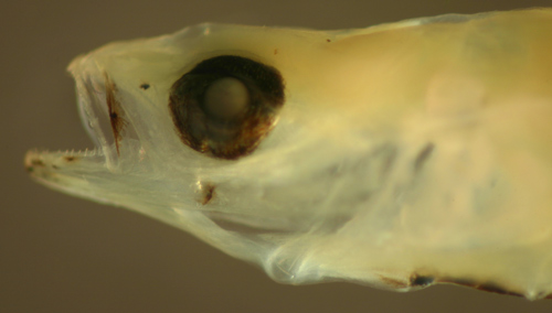
Gobiomorus
dormitor larva
12.1 mm SL
fin-ray count: 10/10
16
San Blas, Panama, SB86-1002
Gobiomorus
dormitor larvae
12.2 and 12.1 mm SL
San Blas, Panama, SB86-1030
Gobiomorus
dormitor larvae
12.2, 12.1 and 12.1
mm SL
San Blas, Panama, SB86-1030
and 1002
Gobiomorus
dormitor larva above
vs. Eleotris amblyopsis larva below
12.1 mm SL
neuromast pattern differences
San Blas, Panama, SB86-1002
Gobiomorus
dormitor transitional recruit
11.3 mm SL
Colon, Panama, N7529a
Erotelis smaragdus
Diagnosis:
Modal fin-ray counts of D-VI,11 A-10 (rarely
10/9) Pect-17 indicate Erotelis smaragdus
or Gobiomorus
dormitor. These genera have overlapping
fin-ray counts, and given the inconsistent ranges
reported in the literature, these cannot be relied
on to distinguish the taxa. Leptophilypnus
fluviatilis, from Panama, may rarely overlap
the dorsal and anal-fin-ray counts (10/10), but
has 18-19 pectoral-fin rays. Larvae and recruits
of Gobiomorus
dormitor differ in having a large eye and
a melanophores only on the first anal-fin membrane.
The larvae of the eastern Pacific sibling species,
Erotelis
armiger, shares the melanophore pattern.
G17
Analogues:
The eleotrid larvae share a long thin body with
relatively short dorsal and anal-fin bases, numerous
procurrent caudal-fin rays, and a long ventral midline
streak from the isthmus to the mid-abdomen. Larval
Erotelis smaragdus are distinguished by a
row of melanophores along the anal-fin membranes
and a characteristic spot on the peduncle midway
along the ventral procurrent caudal-fin rays.
Description:
Body very thin, long and narrow with a medium eye
and a terminal medium-sized mouth. Pelvic fins separate
and long, pectoral fins long, dorsal and anal-fin
bases medium length (the base of the second dorsal
fin can be almost the same length as the caudal
peduncle behind the fin (or at least more than two-thirds)
and the rays are of roughly even length), caudal
peduncle long and relatively narrow and there are
many procurrent caudal-fin rays in a distinct fleshy
fold, typically about 12-14 in this larval type.
Markings mostly along the ventral midline: melanophores
usually as streaks (but sometimes contracted and
appear as spots) extending from the isthmus most
of the way to the vent (about 10), ending at the
swim bladder (which is the full thickness of the
abdomen and provides a clear view of the retroperitoneum).
There is a row of melanophores along the bases of
the fourth and fifth and then the last several anal-fin
elements (variably paired) and continuing as several
streaks along the ventral peduncle ending at the
start of the lower procurrent caudal-fin rays. There
are melanophores on the membranes between all of
the anal-fin elements. There is a gap of melanophores
at the start of the ventral procurrent caudal-fin
rays followed by a patch of melanophores on the
mid procurrent rays and then heavy pigmentation
over the bases of the lower segmented caudal-fin
rays extending along the full length of the lower
segmented caudal rays as well as on the mid and
distal portions of the central and sometimes the
upper segmented caudal-fin rays. The central and
upper portion of the end of the caudal peduncle
is covered by surface melanophores. A small and
often inconspicuous melanophore is frequently present
on the dorsal midline of the caudal peduncle just
forward of the start of the procurrent caudal-fin
rays. Head markings on all individuals include melanophores
at the angle of the jaw, along the dentary at the
tip and below the dentary just to the side of the
tip of the lower jaw. Head markings on some individuals
include melanophores on the inside dentary of the
mid-lower jaw, the rim of the mid-maxilla, the tip
of the upper jaw, and below the nasal bones. Melanophores
extend over the iris on the dorsal half of the eyeball
and around the sides of the base of the eyeball
and, when expanded, melanophores cover most of the
outer half of the iris ring. Many individuals have
a hidden melanophore overlying the cleithrum visible
upon lifting the operculum (variably paired) and
most have a melanophore along the insertion of the
lowest pectoral-fin ray. Internal melanophores surround
the saccule and are present along the dorsal surface
of the swim bladder and paired around the gut near
the vent. There is a linear internal melanophore
above the pelvic girdle, perhaps along the ventral
postcleithrum. There are prominent internal streaks
extending along the hemal spines from the vent level
rearwards (starting at the ventral midline melanophores
and extending upwards toward the vertebral bodies
and then occasionally even above). Series of transitional
larvae show development of the eye from a moderately
narrowed vertical oval, no tilt, with the pupil
off-center dorsally and a posterior-inferior extension
of the iris and/or melanophores (there is often
a marked dorsal and ventral indentation of the iris
as well), to fully round. Transitional larvae develop
a pattern of large discrete melanophores speckling
the upper half of the head, around the nasal bones
and along the upper and lower jaws as well as internally
around the posterior brain case. Surface melanophores
develop along the lateral midline from the caudal
peduncle forwards.
Erotelis
smaragdus larva
11.2 mm SL
San Blas, Panama, SB87-227
Erotelis
smaragdus larva
11.3 mm SL
note internal melanophores
San Blas, Panama, SB86-702
Erotelis
smaragdus larva
11.2 mm SL
note internal melanophores
San Blas, Panama, SB86-826
Erotelis
smaragdus larva
11.4 mm SL
internal melanophores
along hemal spines
San Blas, Panama, SB87-302
Erotelis
smaragdus larva
11.4 mm SL
note iris extension
San Blas, Panama, SB86-1030
Erotelis
smaragdus larva
10.3 mm SL
note indented iris
and
12-14 procurrent caudal-fin rays
San Blas, Panama, SB86-808
Erotelis
smaragdus larva
11.1 mm SL
note melanophores
at cleithrum,
lowest pectoral-fin ray insertion,
and internal pelvic-fin process
San Blas, Panama, SB86-109
Erotelis
smaragdus larvae
transitional series
11.4, 10.8, and 11.3
mm SL
San Blas, Panama, SB86-1030
Erotelis
smaragdus transitional larva
11.3 mm SL
San Blas, Panama, SB86-1030
Erotelis
smaragdus
early transitional larva
10.8 mm SL
San Blas, Panama, SB86-1030
Erotelis
smaragdus transitional recruits
10.7 and 10.9 mm SL
Colon, Panama, N7528b
Erotelis
smaragdus transitional recruit
10.9 mm SL
Colon, Panama, N7528b
Guavina guavina
Diagnosis:
Analogues:
Description:
Leptophilypnus fluviatilis
?
Diagnosis:
A larva with the fin-ray count of D-VI,11
A-10 and numerous procurrent caudal-fin rays (11-12)
could represent any of Erotelis
smaragdus, Gobiomorus
dormitor, Guavina
guavina, or Leptophilypnus fluviatilis.
This larva could possibly represent an earlier-stage
larva of either Gobiomorus
dormitor or Erotelis
smaragdus, however several features support
Leptophilypnus fluviatilis: this larva has
no melanophores on the lateral caudal peduncle (characteristic
of the other two genera, even at smaller sizes);
this larva has 11 procurrent caudal-fin rays but
small immature larvae of the other two genera do
not have that full complement; and, lastly, the
eyeball of the other two genera has a distinctive
ventral extension not present on this larva, perhaps
indicating that this larva is transitional at this
small size. Larval Guavina
guavina are distinctly shorter and wider-bodied.
(PE)
Analogues:
The eleotrid larvae share a long thin body with
relatively short dorsal and anal-fin bases, numerous
procurrent caudal-fin rays, and a long ventral midline
streak from the isthmus to the mid-abdomen. This
larval type resembles larval Dormitator
maculatus with a delicate body and few markings,
but has quite different fin-ray counts, with 11/10
vs. 9/10 and many more procurrent caudal-fin rays.
It has a small and even oval eye, unlike pre-transitional
larval Gobiomorus
dormitor and Erotelis
smaragdus which have a distinctive ventral
extension. It is missing the characteristic patch
of melanophores covering the caudal peduncle of
larval Erotelis
smaragdus and those on the upper half of
the caudal fin of larval Gobiomorus
dormitor. The few true gobies that share
the numerous (>10) procurrent caudal-fin rays
(notably Sicydium
and Awaous
banana) have distinctive internal melanophores
that are absent on this larval type.
Description:
Body thin, long and narrow with a small vertically-narrowed
oval eye and a terminal mouth. Pectoral fins length
unknown, dorsal and anal-fin bases short, pelvic
fins length unknown, caudal peduncle long and relatively
narrow with 11-12 spindly procurrent caudal-fin
rays. Markings mostly along the ventral midline:
melanophores as streaks at the isthmus, the pelvic-fin
insertion, and the mid-abdomen. There are four discrete
melanophores along the base of the anal fin followed
by a streak of melanophores along the ventral peduncle
ending at the first procurrent caudal-fin rays.
Melanophores are present at the base of most of
the lower segmented caudal-fin rays extending well
out along the fin rays. Head markings are limited
to melanophores outlining the lower rim of the dentary
at the tip of the lower jaw. Internal melanophores
are present along the dorsal surface of the swim
bladder.