|
|
|
Group 1: The short-fin gobies
pt. 1 (six-spined, fused)
|
|
Bathygobius, Lophogobius, Priolepis, Awaous, and Sicydium
|
| |
|
This group of six-spined
gobies with short median fins and fused pelvic
fins includes several unrelated genera of gobies,
including tidepool, reef, and fresh-water species.
Although common in their appropriate habitats,
this group of gobies are not usually observed
or photographed on reefs. The abundant reef and
sand gobies of Coryphopterus
and Lythrypnus are
separated for convenience and treated in Group
2.
|
|
|
Note: Fin-ray counts for the second dorsal fin and
the anal fin are total elements (spines plus rays)
and species are listed in rough order of increasing
fin-ray counts. |
|
|
|
|
|
|
|
|
|
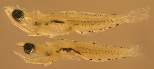
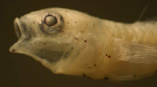
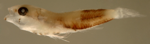
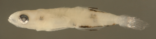
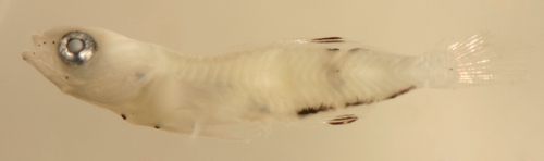
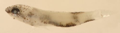
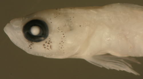
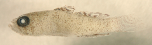
Diagnosis:
Modal fin-ray counts of D-VI,10 A-9 and Pect-15-17
with fused pelvic fins indicate Bathygobius
curacao and Lythrypnus (and
overlaps the range of Coryphopterus alloides).
These genera typically have one fewer anal-fin
ray than second-dorsal-fin rays. Larval Lythrypnus are
lightly-marked and develop radiating bars of melanophores
around the eye at transition. Coryphopterus
also have lightly-marked larvae and C. alloides
is the only species which would overlap this fin-ray
count, although only rarely with 15 pectoral-fin
rays. Lophogobius cyprinoides
and Priolepis hipoliti share the
median-fin ray count but have more pectoral-fin
rays. Bathygobius are known for having
the dorsal-most pectoral-fin rays separate from
the rest and filamentous, however this feature
is not apparent on larvae. This larval type has
15-17 pectoral-fin rays, indicating the species
is B. curacao (B. soporator and B. mystacium have a mode of
19-20 pectoral-fin rays). (DNA) G14a
|
|
| Analogues:
(heavy ventral markings) |
|
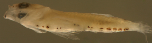
| Description:
Body relatively thin, long and narrow with a large
eye and a terminal large mouth. Pectoral fins long,
reaching to vent. Pelvic fins long, reaching almost
to the vent, with an obvious pelvic frenum. Dorsal
and anal-fin bases medium-length and caudal peduncle
medium-length and sharply narrowing, 7-9 procurrent
caudal-fin rays (7-8 spindly). Heavily marked; along
the ventral midline there are large point, stellate,
or streak melanophores at the isthmus, one or two
forward of the pelvic-fin insertion, and a few behind
the pelvic-fin insertion along the abdominal midline.
Then there is a variable row of two or three large
paired melanophores spaced along the anal-fin base,
continuing as a row of three or four large single
melanophores along the caudal peduncle ending at
the start of the procurrent caudal-fin rays (the
anal-fin base and caudal peduncle melanophores often
merge into a single long streak). Dorsal markings
consist of a row of paired melanophores on either
side of the dorsal midline: a pair just forward
of the spinous dorsal fin, one just behind, then
two or three pairs spaced along the soft dorsal
fin, followed by one to three unpaired melanophores
along the dorsal midline of the caudal peduncle
ending well before the start of the upper procurrent
caudal-fin rays. Markings on the head consist of
a large melanophore outlining the lower edge of
the dentary at the tip of the lower jaw and another
at the angle of the jaw (on pre-transitional larvae).
Internal melanophores are present at the base of
the braincase (sometimes around the upper braincase
as well), at the sacculus, along the dorsal surface
of the peritoneum and swim bladder, and continuing
along the gut to the vent (often all of these merge
into a dark streak arcing through the body). There
is a row of internal vertebral melanophores above
and usually below the vertebral bodies from the
mid-body to the caudal peduncle. This streak can
be prominent or mostly obscured by overlying musculature.
Some individuals have melanophores at the base of
the lower segmented caudal-fin rays extending out
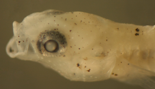
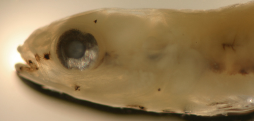
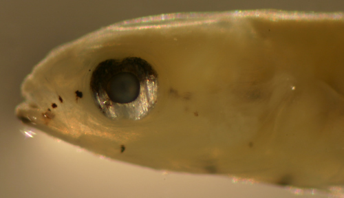
a short distance along the rays. Series of transitional
larvae show development of the eye from round with
dorsal and ventral indentations in the iris (mostly
on the dorsal-anterior to ventral-posterior axis,
but can vary) to fully round (most pre-transitional
larvae captured have no indentations, and some transitional
larvae have iris indentations). Transitional larvae
intensify the surface melanophores on the iris (covering
the upper third of the eyeball and at 2, 5 and 7-8
o'clock) and develop a stripe from the eye forward
across the mid-upper jaw to the mid-lower jaw and
a stripe of melanophores behind the eye across the
mid-operculum. Transitional larvae then develop
a speckling of large melanophores and leukophores
on the top of the head to the base of the pectoral
fin and a stripe of iridophores across the operculum
and onto the base of the pectoral fin. |
|
|
| |
|
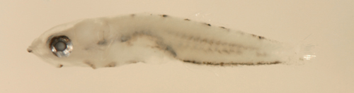
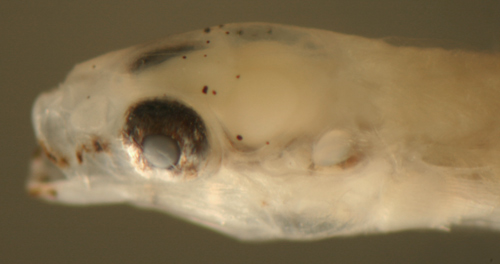
| Bathygobius
curacao larva |
| 5.2 mm SL |
| melanophores in streaks
|
| San Blas, Panama, SB86-426 |
| |
|
 |
| |
 |
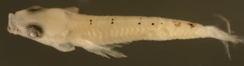
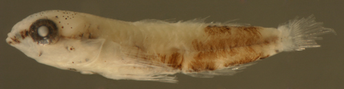
| Bathygobius
curacao larva |
| 6.8 mm SL |
| San Blas, Panama, SB87-225 |
| |
|
 |
| |
 |
| |
 |
| |
 |
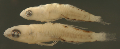
| Bathygobius
curacao larvae |
| 4.7 and 5.9 mm SL |
| smallest larva above,
size comparison |
| San Blas, Panama, SB86-1010 |
| |
|
 |
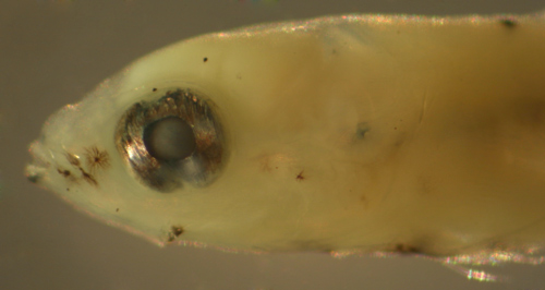
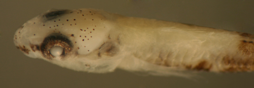
| Bathygobius
curacao transitional larva |
| 5.5 mm SL |
| with iris indentations |
| San Blas, Panama, SB87-219 |
| |
|
 |
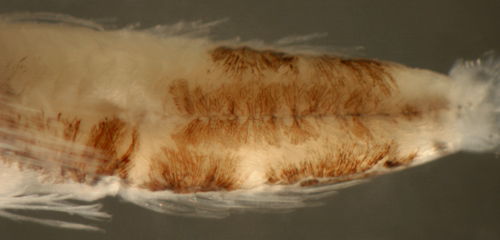
| Bathygobius
curacao transitional larvae |
| 5.3 and 4.9 mm SL |
| internal melanophores |
| San Blas, Panama, SB86-1123 |
| |
|
 |
| Bathygobius
curacao transitional series |
| 5.3, 5.4, and 5.9 mm
SL |
| San Blas, Panama, SB86-426 |
| |
|
 |
| Bathygobius
curacao transitional larva |
| 6.0 mm SL |
| note head neuromasts |
| San Blas, Panama, SB86-1010 |
| |
|
 |
| |
 |
| |
 |
|
|
|
|
|
|
|
|
|
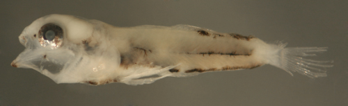
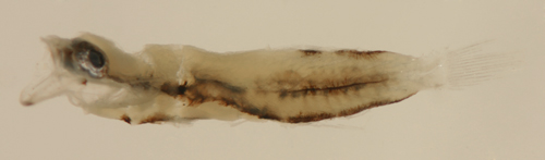
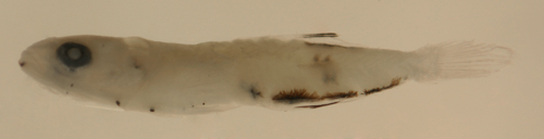
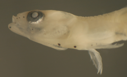
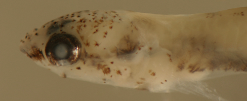
Diagnosis:
Modal fin-ray counts of D-VI,10 A-9 and Pect-19
with fused pelvic fins indicate Bathygobius
mystacium or B. soporator. The two species
are usually separated by the former having 35
(33-36) and the latter 37-41 scale rows later
in development. The genus is known for having
the dorsal-most pectoral-fin rays separate from
the rest and filamentous, however this feature
is not apparent on larvae. This larval type has
a mode of 19 pectoral-fin rays, consistent with
either B. soporator or B. mystacium.
Since DNA sequences match the "spot"
type Bathygobius larva to B.
soporator, this larval type matches B.
mystacium. Other six-dorsal-spined gobies
with the same median-fin ray counts (but fewer
pectoral-fin rays) include the congener B. curacao with 16-17, as well
as Lythrypnus
(14-16), Coryphopterus
alloides (16-17), Lophogobius
cyprinoides (17-18), and Priolepis
hipoliti (18). (ML) G14b
|
|
| Analogues:
(heavy ventral markings) |
|
| Description:
Body relatively thin, long and narrow with
a large eye and a terminal mouth. Pectoral fins
long, reaching to the vent. Pelvic fins long, reaching
almost to the vent, with an obvious pelvic frenum.
Dorsal and anal-fin bases medium-length and caudal
peduncle medium-length and sharply narrowing, 7-9
procurrent caudal-fin rays (7-8 spindly). Heavily
marked mostly along the lower and midbody with markedly
dendritic melanophores: there is a large melanophore
at the tip of the lower jaw and at the angle of
the jaw. Along the ventral midline there are large
stellate or streak melanophores at the isthmus,
forward and behind of the pelvic-fin insertion,
then a variable row (paired, one per side) at the
anal-fin base and then unpaired extending along
the caudal peduncle ending at the start of the procurrent
caudal-fin rays. Internal melanophores occur around
the lower brain case and around the sacculus continuing
along the dorsal surface of the peritoneum and swim
bladder extending to the gut near the vent (often
all of these merge into a dark streak arcing through
the body). There is a row of internal melanophores
surrounding the vertebral bodies and extending for
most of the spine from the level of the vent to
the mid-caudal peduncle, often with a discrete row
of deep melanophores along the dorsal vertebral
spines as well. There is a prominent and characteristic
matching row of dendritic surface melanophores along
the lateral midline. Melanophores along the dorsal
midline are limited to the rear body (vs. B. curacao ), as two or three
variably-paired large stellate melanophores on either
side of the dorsal midline at the base of the mid
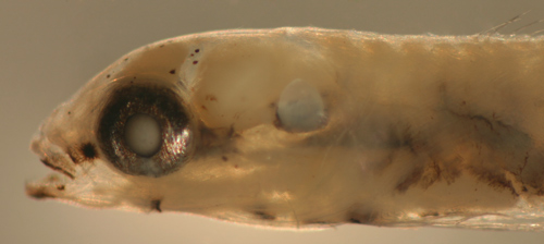
to rear soft dorsal fin. Series of transitional
larvae show development of the eye from round with
dorsal and ventral indentations in the iris (mostly
on the dorsal-anterior to ventral-posterior axis,
but can vary) to fully round (most pre-transitional
larvae captured have no indentations, and some transitional
larvae have iris indentations).with melanophores
extending in patches across the surface of the iris.
Transitional larvae develop a stripe of melanophores
from the eye forward to the mid-upper jaw and across
to the mid-lower jaw. As transition continues, the
melanophores become essentially a stripe from the
tip of the lower jaw back across the mid-upper jaw
to the eye, over the iris, continuing internally
over the base of the braincase to the sacculus continuing
internally to the dorsal surface of the swim bladder
then to the vertebral row of melanophores. A branch
stripe extends bilaterally along the internal lateral
abdominal wall to the vent and along the base of
the anal fin to the tail. A scattering of large
melanophores and some leukophores develops on the
top of the head. |
|
|
| |
|
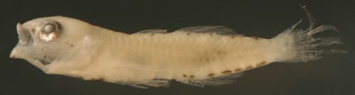
| Bathygobius
mystacium larva |
| 5.9 mm SL |
| San Blas, Panama, SB86-425 |
| |
|
 |
| |
 |
| |
 |
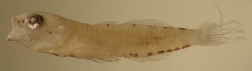
| Bathygobius
mystacium larva |
| 5.7 mm SL |
| San Blas, Panama, SB86-808 |
| |
|
 |
| |
 |
| Bathygobius
mystacium larva |
| 5.9 mm SL |
| with iris indentations |
| San Blas, Panama, SB87-218 |
| |
|
 |
| Bathygobius
mystacium larva |
| 5.3 mm SL |
| internal melanophores
|
| San Blas, Panama, SB84-523 |
| |
|
 |
| Bathygobius
mystacium transitional larva |
| 6.3 mm SL |
| internal melanophores
|
| San Blas, Panama, SB87-219 |
|
|
 |
| Bathygobius
mystacium transitional larva |
| 5.8 mm SL |
| San Blas, Panama, SB86-808 |
| |
|
 |
| Bathygobius
mystacium transitional larva |
| 5.9 mm SL |
| San Blas, Panama, SB86-1008 |
| |
|
 |
| |
 |
| |
 |
|
|
|
|
|
|
|
|
|
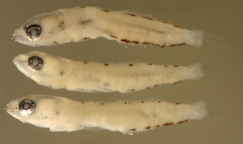
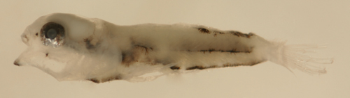
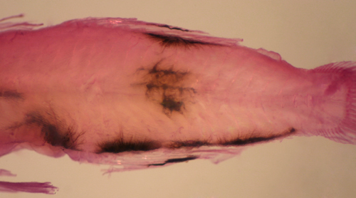
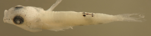
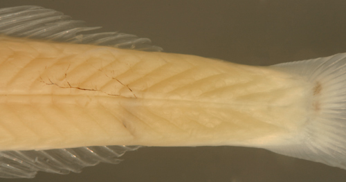
Diagnosis:
Modal fin-ray counts of D-VI,10 A-9 and Pect-19
with fused pelvic fins indicate Bathygobius
soporator or B. mystacium. The two species
are usually separated by the former having 37-41
and the latter 35 (33-36) scale rows later in
development. The genus is known for having the
dorsal-most pectoral-fin rays separate from the
rest and filamentous, however this feature is
clearly not apparent on larvae. The DNA sequence
of this larval type (the "spot" type,
i.e. a single large vertebral melanophore) matches
adult B. soporator. Other six-dorsal-spined
gobies with the same median-fin ray counts (but
fewer pectoral-fin rays) include the congener
B. curacao with 16-17, as well
as
Lythrypnus
(14-16), Coryphopterus
alloides (16-17), Lophogobius
cyprinoides (17-18), and Priolepis
hipoliti (18). (DNA) G14
|
|
| Analogues:
(heavy ventral markings) |
|
| Description:
Body relatively thin, long and narrow with
a large eye and a terminal mouth. Pectoral and pelvic
fins long, reaching almost to the vent, with a obvious
pelvic frenum. Dorsal and anal-fin bases medium-length
and caudal peduncle medium-length and sharply narrowing,
7-9 procurrent caudal-fin rays (7-8 spindly). Heavily
marked mostly along the lower and midbody: there
is a large melanophore at the tip of the lower jaw
and one at the angle of the jaw. Along the ventral
midline there are large stellate or streak melanophores
at the isthmus, the pelvic-fin insertion, and one
to three along the mid-abdomen, then variably paired
on either side of the ventral midline at the anal-fin
base and then extending along the ventral peduncle
ending at the start of the procurrent caudal-fin
rays. Internal melanophores occur around the sacculus
and along the dorsal surface of the swim bladder
and around the gut near the vent. Melanophores along
the dorsal midline are limited to the rear body
(vs. B. curacao); as one to three
variably paired large stellate melanophores on either
side of the dorsal midline at the base of the mid
to rear soft dorsal fin. Long streak melanophores
are present along the membranes of the second to
fifth fin rays on both the soft dorsal and anal
fins. There is a single prominent stellate internal
vertebral melanophore at the lateral midline at
about the level of the mid soft dorsal fin that
ramifies around the vertebral bodies and extends
between and around myomeres and often up to the
surface. Series of transitional larvae show development
of the eye from round with dorsal and ventral indentations
in the iris (mostly on the dorsal-anterior to ventral-posterior
axis, but can vary) to fully round (most pre-transitional
larvae captured have no indentations and some transitional
larvae have iris indentations). Early transitional
larvae develop a stripe of melanophores from the
eye forward to the mid-upper jaw. As transition
continues, the melanophores become essentially a
stripe from the tip of the lower jaw across the
mid-upper jaw to the eye, over the iris and onto
the operculum, continuing internally from the sacculus
to the dorsal surface of the swim bladder and around
the gut near the vent and along the anal fin to
the tail. Melanophores also develop at the end of
the caudal peduncle, primarily at the base of the
central and lower segmented caudal-fin rays. Series
of transitional larvae show the eye remaining round,
but becoming larger with the iris developing a dark
surface pigmentation layer. Late transitional larvae
develop an additional scattering of large discrete
melanophores on the dorsal half of the head and
the operculum, extending to the base of the pectoral
fin. Small iridophores occur in patches behind the
eye and in a stripe out onto the middle rays of
the pectoral fin. Patches of small melanophores
develop around the base of the spinous dorsal fin. |
|
|
| |
|
| Bathygobius
soporator larva |
| 5.5 mm SL |
| San Blas, Panama, SB86-425 |
| |
|
 |
| |
 |
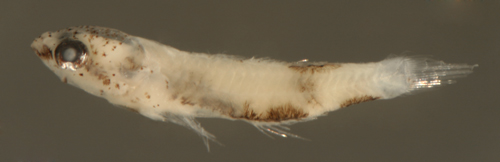
| Bathygobius
soporator larva |
| 5.6 mm SL |
| single branching vertebral
melanophore |
| San Blas, Panama, SB86-425 |
| |
|
 |
| Bathygobius
soporator larva |
| 5.7 mm SL |
| with iris indentations |
| San Blas, Panama, SB87-218 |
| |
|
 |
| Bathygobius
soporator early transitional |
| 6.0 mm SL |
| San Blas, Panama, SB86-425 |
| |
|
 |
| |
 |
| |
 |
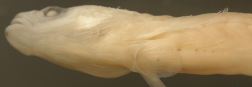
| Bathygobius
soporator transitional larva |
| 5.6 mm SL |
| note pelvic-fin frenum |
| San Blas, Panama, SB86-616 |
| |
|
 |
| |
 |
| Bathygobius
soporator transitional larva |
| 5.8 mm SL |
| San Blas, Panama, SB86-426 |
| |
|
 |
| |
 |
| |
 |
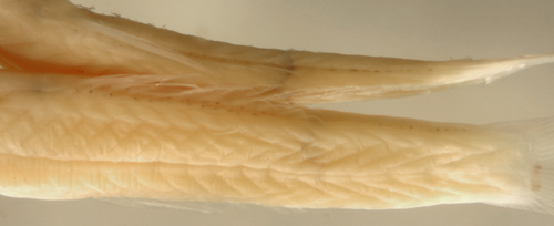
| Bathygobius
soporator transitional recruit |
| 7.4 mm SL |
larval melanophore
remnants
on dorsal and anal-fin ray membranes |
| Noronha, Brazil FN01 |
| |
|
 |
| |
 |
|
|
|
|
|
|
|
|
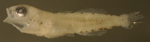
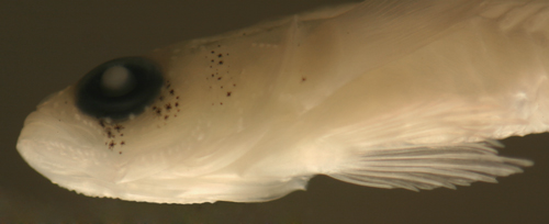
| Diagnosis:
Modal fin-ray counts of D-VI,10 A-9 and Pect-18
are shared by Lophogobius cyprinoides and
Priolepis
hipoliti. L. cyprinoides has a pelvic
frenum, which is absent in larval and juvenile P.
hipoliti. Coryphopterus
alloides matches the median-fin ray counts
but has fewer pectoral-fin rays (16-17) and recruits
are pale sand gobies with a prominent internal dark
mid-body bar. Bathygobius
mystacium and B.
soporator have more pectoral-fin rays (mode
of 19-20). B.
curacao and Lythrypnus
have fewer pectoral-fin rays (15-17 and 14-16). |
|
| Analogues:
(VMS4: jaw angle, thorax, anal fin, caudal peduncle)
The larval stage has not been identified for Lophogobius
cyprinoides, however, based on the transitional
recruit, the melanophore pattern would be similar
to the Bathygobius,
Lythrypnus,
and Coryphopterus
larval types. Unfortunately, the latter taxa are
very common and diverse in larval collections, making
it possible that the larvae of L. cyprinoides
may have been subsumed in those. Fin-ray counts
do differ, but only slightly. Notably, if it is
consistent, the absence of a second thoracic melanophore
anterior to the pelvic-fin insertion would be important
since these other larval types have two thoracic
midline melanophores. Bathygobius
larvae have either fewer or more pectoral-fin
rays and distinctive internal melanophores not obviously
apparent on the transitional L. cyprinoides.
Larval Coryphopterus
only rarely have 10/9 median fin elements;
the one species with that count, C.
alloides, has fewer pectoral-fin rays and
more procurrent caudal-fin rays. Lythrypnus
have fewer pectoral-fin rays. The seven-spined
gobies with similar larvae do not have the jaw
angle melanophores and the caudal peduncle streak
extends only halfway to the caudal fin. The seven-spined
Barbulifer
larvae share the median fin-ray count and
the jaw angle melanophores, but have additional
melanophores and a flattened head. |
|
| Description:
Based on the transitional recruit, the body is somewhat
long and narrow (although wider anteriorly than
most goby larvae) with a large eye and a terminal
mouth. Pectoral fins long, pelvic fins long and
fused with an obvious pelvic frenum, caudal-fin
procurrent rays 6-7 (6 spindly). Dorsal and anal-fin
bases relatively short, caudal peduncle rapidly
narrowing. Larval melanophores apparent on the transitional
recruit include those at the jaw angle and a series
along the ventral midline: a single one at the thorax,
a row along the base of the anal-fin rays, and streaks
along the caudal peduncle extending up to the procurrent
caudal-fin rays. |
|
|
|
| Lophogobius
cyprinoides |
| transitional recruit |
| 7.4 mm SL |
| Colon, Panama, N7527b |
|
 |
|
|
 |
| Lophogobius
cyprinoides recruit |
| 9.3 mm SL |
| Colon, Panama, N7527b |
|
 |
|
|
 |
| |
|
|
|
|
|
|
|
|
|
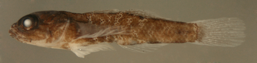
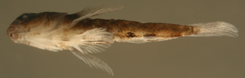
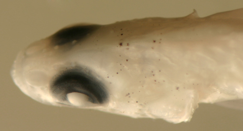
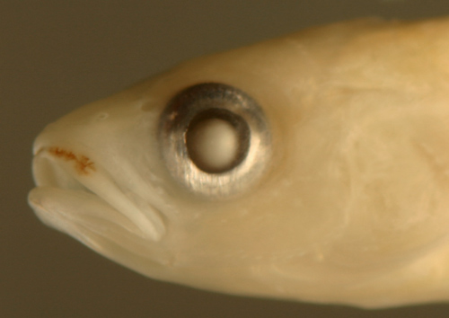
| Diagnosis:
Modal fin-ray counts of D-VI,10 A-9 and Pect-18
indicate Priolepis hipoliti and Lophogobius
cyprinoides. P. hipoliti larvae and
juveniles are missing the pelvic frenum, present
on L.
cyprinoides and most other gobies. Priolepis
robinsi from Colombia and the Brazilian P.
dawsoni have higher median-fin ray counts (D-VI,11
A-10). Coryphopterus
alloides match the median-fin ray counts
but have fewer pectoral-fin rays (16-17) and recruits
have a prominent internal dark mid-body bar. Coryphopterus
kuna has 9/9 and pect. 15. Bathygobius
mystacium and B.
soporator have more pectoral-fin rays (mode
of 19-20). B.
curacao and Lythrypnus
have fewer pectoral-fin rays (15-17 and
14-16). (DNA) |
|
| Analogues:
|
|
| Description:
Body long and narrow with a large eye and
a terminal large mouth. Pectoral fins long, pelvic
fins long and fused with clearly no pelvic frenum.
Dorsal and anal-fin bases relatively short, caudal
peduncle long and rapidly narrowing. The first dorsal
and anal-fin rays are long and the last short, making
a somewhat triangular fin shape. Series of transitional
larvae show the eye remains round but the head thickens
and the body hunches over. Transitional larvae develop
melanophores in bars below the eye and an arc across
the top of the head behind the eye and down the
preopercle. Sensory papillae develop in rows on
the head. Transitional recruits show a pattern of
dark median fins and bars on the head and body. |
|
|
|
| Priolepis
hipoliti transitional larva |
| 9.8 mm SL, DNA confirmed
ID |
| Belize, BCN52, coll.
by C. Nolan |
|
 |
| |
 |
| |
 |
| |
 |
| Priolepis
dawsoni recruit |
| 10.1 mm SL |
| Noronha, Brazil, FN01 |
|
 |
| |
 |
| |
|
|
|
|
|
|
|
|
|
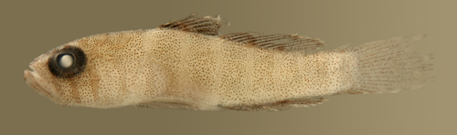
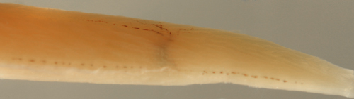
| Diagnosis:
Long thin larvae with a modal fin-ray count of D-VI,11
A-11 Pect-16 indicates the river goby Awaous
banana. Coryphopterus
personatus shares the fin-ray counts but
their larvae are very different; they are smaller
and shorter and have typical ventral midline melanophore
rows and no large internal melanophores. A. flavus
from Colombia to Brazil has a modal fin-ray
count of D-VI,10 A-10 Pect-16. |
|
| Analogues:
Compared to other long thin goby larvae, Awaous
and Sicydium
larvae have shorter dorsal and anal fins and many
more procurrent caudal-fin rays. The gobioid sleeper
family, the eleotrids,
share these two attributes, but have clearly-divided
pelvic fins and lack the large internal melanophore
over the rear end of the anal fin. The larvae of
Sicydium
are quite similar to Awaous banana
in form, markings, and median-fin ray counts. The
primary meristic difference is higher pectoral fin-ray
counts in larval Sicydium
(20 or more). Both sets of larvae share the large
internal melanophore over the anal fin, but Sicydium
larvae have additional melanophores, in
particular at the base of the upper caudal fin,
deep to the pectoral-fin base, and along the mid-abdominal
ventral midline. In addition, the large internal
melanophore in larval Sicydium
has characteristic, sometimes extreme, long filamentous
extensions. |
|
| Description:
Body thin, long, and narrow with a medium-sized
round eye and a terminal small mouth. Pectoral fins
very short, pelvic fins very short. Dorsal and anal-fin
bases short and caudal peduncle sharply narrowing,
10-14 procurrent caudal-fin rays. Melanophores on
the head only along the dorsal edge of the anterior
premaxilla on each side and midline near the tip
of the lower jaw. Ventral melanophores are limited
to a paired large melanophore at the mid-base of
the anal fin and internal melanophores overlying
the posterior swim bladder and extending down to
the vent. A large deep internal vertical melanophore
underlies the last anal-fin ray extending up to
the lateral midline. |
|
|
|
| Awaous
banana larva |
| 12.3 mm SL, DNA confirmed
ID |
| Yucatan, Mexico, 240306 |
| coll. by Lourdes Vasquez
et al. |
|
 |
|
|
 |
|
|
 |
|
|
 |
| |
|
|
|
|
|
|
|
|
|
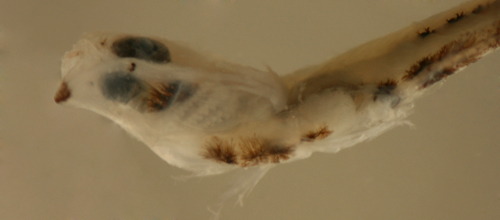
| Diagnosis:
Long thin larvae with a short round pelvic-fin sucking
disk and modal fin-ray counts of D-VI,11 A-11 Pect-20
indicate the river gobies of Sicydium. The
taxonomy and range of species in this genus are
generally unresolved and numerous species have been
described. I follow the recent review for Central
America by Lyons (2005). He indicates that S.
altum ranges along the coast from Costa Rica
through Panama to Colombia, while S. adelum is
localized to Costa Rica and S. plumieri and
S. punctatum occur in the far east of Panama
near Colombia and probably beyond. The remaining
Central American species is the northern Sicydium
gymnogaster, found only from Veracruz, Mexico
to Honduras. There are many other named Caribbean
species, some with very restricted ranges, including
S. vincente (West Indies), S. caguitae
(Puerto Rico), S. montanum (Venezuela), S.
buscki and S. gilberti (both Dominican
Republic), while S. antillarum is
considered widespread. |
|
| Analogues:
|
|
Description:
Body thin, long, and narrow with a medium-sized
round eye and a terminal small mouth. Pectoral fins
very short, pelvic fins very short. Dorsal and anal-fin
bases short and caudal peduncle long and narrowing,
10-14 procurrent caudal-fin rays (this large number
shared only with some of the eleotrids).
Immature larvae long and thin with Melanophores
on the head only along the dorsal edge of the front
premaxilla on each side and one midline below the
dentary at the tip of the lower jaw. There are ventral
midline Internal melanophores are present at the
dorsal surface of the swim bladder and around the
gut near the vent. melanophores at the base of the
upper segmented caudal-fin rays (an unusual pattern
for gobiid larvae). There is a large and characteristic
internal melanophore at the mid-body above the anal
fin that extends in long dendritic filaments up
to the surface and along the skin (usually towards
the head).
Mature larvae are large and stout with a rounded
cross-section. The pelvic fins are very short and
rounded with an obvious frenum. Mature larvae retain
the melanophore patterns of immature larvae, but
the melanophores along the ventral midline are present
at the pelvic-fin insertion and behind the pelvic-fin
insertion, followed by a short row of melanophores
along the lateral wall of the abdomen on each side.
There is a row of melanophores along the anal-fin
base (variably paired, one per side, more than one
per fin ray) and then extending unpaired along the
ventral midline of the caudal peduncle ending near
the start of the procurrent caudal-fin rays. The
melanophores at the base of the upper segmented
caudal-fin rays persist and the large internal melanophore
remains prominent. |
|
|
|
| Sicydium
altum larva |
| 25.7 mm SL |
| San Blas, Panama,
SB80-101 |
|
 |
| |
 |
| Sicydium
altum larva |
| 23.2 mm SL |
| San Blas, Panama,
SB86-702 |
|
 |
| Sicydium
altum larva |
| 23.8 mm SL |
| San Blas, Panama,
SB80-101 |
|
 |
| Sicydium
altum larvae |
| 23.8 mm SL (above)
25.7 mm SL (below) |
| San Blas, Panama,
SB80-101 |
|
 |
| |
 |
| Sicydium
altum larvae |
| 23.8 mm SL (above)
25.7 mm SL (below) |
| San Blas, Panama,
SB80-101 |
|
|
|
| |
|
|
|
|
|
|
| Sicydium gymnogaster/plumieri |
|
|
|
|
|
|
|
|
|
| Diagnosis:
Long thin larvae with a short round pelvic-fin sucking
disk and modal fin-ray counts of D-VI,11 A-11 Pect-20
indicate the river gobies of Sicydium. The
taxonomy and range of species in this genus are
generally unresolved and numerous species have been
described (Bussing 1995). According to Lyons (2005)
the northern Central American coastal species is
S. gymnogaster, found from Veracruz, Mexico
to Honduras, but this Yucatan-caught larvae is a
DNA match to an adult from St. Thomas USVI, indicating
that the species limits and/or phylogenetics are
in question. |
|
| Analogues:
|
|
|
Description: Body
thin, long, and narrow with a medium-sized round
eye and a terminal small mouth. Pectoral fins
very short, pelvic fins very short. Dorsal and
anal-fin bases short and caudal peduncle narrowing,
10-14 procurrent caudal-fin rays. On the head
there are melanophores lining the premaxilla and
the dentary at the tip of the lower jaw and a
pair of large melanophores at the rear edge of
the brain case. A large sub-surface melanophore
is placed behind the pectoral-fin base on each
side of the body. Along the ventral midline there
is a large melanophore at the mid-abdomen and
then paired at the base of the mid-anal-fin. Internal
melanophores overlie the posterior swim bladder
and extend down to the vent. A large deep internal
vertical melanophore underlies the last anal-fin
ray extending to the lateral midline, where it
surfaces and spreads as a large dendritic surface
melanophore with characteristically long filamentous
extensions. Melanophores are concentrated at the
base of the mid and upper caudal-fin segmented
rays.
|
|
|
|
| Sicydium
gymnogaster larva |
| 11.8 mm SL, DNA confirmed
ID |
| Yucatan, Mexico, 200306 |
| coll. by Lourdes Vasquez
et al. |
|
 |
|
|
 |
|
|
 |
| |
|
|
|
|
|
 |
|
All contents © copyright 2006-2013
All rights reserved
www.coralreeffish.com by Benjamin
Victor
|
|
|
|
|