|
|
|
| INTRODUCTION
TO LUTJANID LARVAE |
| The
snappers are a prominent family of predatory
fishes found in all tropical waters and often
associated with reef or mangrove habitats.
The deeper-water species, in particular, are
an important component of fisheries in the
Caribbean and the Gulf of Mexico. There are
numerous snapper species in the region and
most of them can be found on or near coral
reefs. The shallow-water snappers are almost
all members of the large genus
Lutjanus. The one exception, the
Yellowtail Snapper Ocyurus
chrysurus, falls well within the Lutjanus
clade in phylogenetic studies. There
are four additional deep-water lutjanid genera,
three of which have only a single Caribbean
representative. |
| |
| Larval
snappers exhibit the standard percoid characters
shared to some degree by many other families:
a wide body, large round eye, large terminal
mouth, stout spines in the fins and a short
anal fin with three spines. Later-stage lutjanid
larvae are distinctive in having a large non-serrated
spine at the angle of the preopercle. Fin-ray
counts broadly overlap with two other common
reef-fish families: the seabasses and groupers
(Serranidae)
and the grunts (Haemulidae).
Separating larval snappers from serranids
is not always easy and useful characters are
discussed in detail below. Grunts are easy
to distinguish since they have narrower bodies,
short non-serrated dorsal-fin spines, two
anal-fin spines (as larvae and small juveniles),
and are typically much smaller at each stage
of development. |
| |
| Regional
Species: There are at least ten
Lutjanus species in the region.
The shallow-water species include a pair of
small snappers with a lateral spot: the Mahogany
Snapper,
L. mahogoni, and the Lane Snapper,
L. synagris. This pair is distinct
in having only twelve dorsal-fin soft rays
(D-X,12); the remaining species in the genus
have fourteen (D-X,14). The Mutton Snapper
L. analis also has a lateral spot.
The remaining shallow-water species have bars
and/or a stripe through the eye and comprise
the Schoolmaster Snapper, L.
apodus, and the Dog Snapper, L.
jocu, which appear similar and differ
primarily in lateral-line scale counts and
some markings. The Gray Snapper, L.
griseus, and the Cubera Snapper, L.
cyanopterus, also look similar, but
Cubera Snappers grow to a much larger size,
up to 125 pounds and over four feet in length.
My mtDNA sequence analyses indicate that similarity
in appearance does not always reflect relatedness:
Gray Snappers are part of the Schoolmaster/Dog
clade while the Cubera Snapper is well out
on its own branch in the phylogenetic tree.
The snappers with a lateral spot do all share
a lineage, but the Yellowtail Snapper Ocyurus
chrysurus, falls in the middle of
that grouping, despite having no spot and
a different morphology. |
| |
| Three
Lutjanus species are predominantly
deep-water denizens, although juveniles occasionally
occur in shallower reef areas: the Blackfin
Snapper L.
buccanella, the Silk Snapper L.
vivanus, and the Red Snapper, L.
campechanus. The Southern Red Snapper,
L. purpureus,
has recently been shown to have identical
DNA sequences at multiple loci to the Red
Snapper, L. campechanus,
confirming that it represents the southern
population of the Red Snapper (Gomes et al.
2008). L. campechanus
from the Gulf of Mexico typically have nine
anal-fin rays, but outside of the Gulf they
mostly share the modal eight anal-fin rays
of the other species in the genus. These deep-water
Lutjanus have typical fin-ray counts
for the genus (D-X,14 Pect-17) and, in genetic
studies, fall within the same clade as the
lateral-spot snappers. Interestingly, they
also have lateral spots to some degree as
juveniles. |
| |
| There
are four other lutjanid genera with a single
Caribbean species each. The common Yellowtail
Snapper Ocyurus
chrysurus (D-X,13 A-III,9 Pect-15-16)
forages in open water and thus has a more
streamlined body form than other snappers
but falls well within the
Lutjanus clade. Three deep-water
snapper genera have a single regional species
each and comprise
Apsilus dentatus (D-X,10 Pect-15-16),
Etelis oculatus
(D-X,11 Pect-15-16), and Rhomboplites
aurorubens (D-XII,11 Pect-17-18). The
remaining deep-water snapper genus has three
species, all with the same fin-ray counts
of D-X,11 Pect-15-16: i.e.
Pristipomoides aquilonaris, P.
freemani, and P.
macrophthalmus. |
| |
Diagnostic
Characters for Lutjanid Larvae:
|
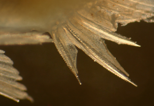
| Early-stage
Larvae: Less-developed lutjanid larvae
occasionally appear in collections made over
the reef. They conform to many of the basic
features of percoid larvae and have a large
head, large round eye, large mouth with a
prominent jaw angle, prominent preopercular
spines, a wide body, continuous dorsal fins
with stout spines, a short anal fin with three
stout spines, and elongated and stout pelvic-fin
spines. Features more specific to lutjanids,
especially Lutjanus, are the moderately-serrated
dorsal and pelvic-fin spines (these spines
and the anal-fin spines often have anterior
serrations as well), the first pelvic-fin
ray longer than the spine, a non-serrated
spine at the angle of the preopercle, and,
most distinctive, a post-cleithral spine. |
| |
| A
number of families have similar-appearing
early-stage larvae, fortunately few occur
in the Atlantic. The most likely confusion
in the region is with serranid
larvae, especially since there is some overlap
in fin-ray counts between Caribbean lutjanid
and serranid species. In general, serranid
species in the region have only seven (serranines)
or nine or ten (epinephelines)
anal-fin soft rays, while most lutjanids have
eight, but this is a fine point for distinguishing
larvae. |
| |
| The
most difficult to distinguish at early stages
are the D-X,12 snappers and the Serranus
species (the epinepheline
serranids have more dorsal-fin soft rays).
In this case, the early-stage larval snappers
have mildly-serrated fin-ray spines while
the Serranus
have smooth spines. Interestingly, these two
unrelated taxa can have quite similar basic
melanophore patterns, however the Serranus
all have a melanophore at the angle of the
jaw and, if intact, obvious speckling of the
pectoral fin-ray membranes. |
| |
| There
is a slight fin-ray-count difference between
the D-X,14 snappers and the epinepheline
serranids. Virtually all of the Caribbean
epinephelines have either eight, nine, or
eleven dorsal-fin spines and most have more
than 14 dorsal-fin soft rays and nine or ten
anal-fin soft rays, while lutjanids have ten
dorsal-fin spines (one with 12), 14 or fewer
dorsal-fin soft rays, and eight anal-fin soft
rays (two with nine). In addition, the few
overlapping epinephelines have 18 or more
pectoral-fin rays (vs. 16-17 in the lutjanids). |
| |
| The
pretransitional larval epinephelines
have markedly-serrated and extended dorsal
and pelvic-fin spines, while the snappers
do not. In addition, the second dorsal-fin
spine is usually much longer than the third
vs. slightly longer or similar in length in
the late-stage larval snappers (this distinction
may not apply to some deep-water snapper genera).
The snapper preopercular spine is notably
non-serrated, while that of the epinephelines
is serrated, but that is not always obvious
on initial inspection. In addition, lutjanids
have the first pelvic-fin soft ray longer
than the spine vs. distinctly shorter in epinephelines.
Finally, there is a characteristic post-cleithral
spine in lutjanid larvae that is not present
in the serranids. (Note: special thanks to
Jeff
Leis and his books). |
|
|
|
|
|
|
|
|
|
|
|
|
|
|
|
|
|
|
|
|
|
|
|
|
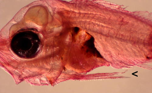
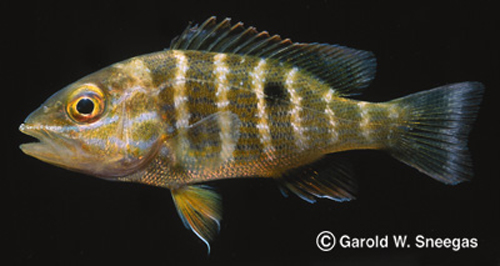
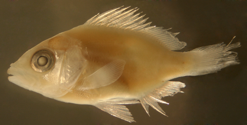
| Lutjanus
campechanus larva |
10 days post-hatch,
5.0 mm TL
laboratory-raised by Jason Lemus and Angelos
Apeitos, Mississippi, USA |
|
 |
 |
| Lutjanus
campechanus larval otoliths |
10 days post-hatch,
5.0 mm TL
lapillus (left), sagitta (right)
bars 100 microns apart |
|
 |
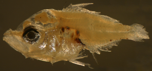
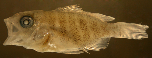
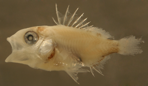
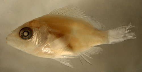
| Lutjanus
(synagris) larva |
| 5.1 mm SL |
Dorsal formula X,12
note post-cleithral spine and serrated
pelvic-fin spines (vs. Serranus),
and first pelvic-fin ray longer than spine
(vs. the epinepheline
groupers ) |
| San Blas, Panama, SB86-1103 |
|
 |
| |
 |
| |
 |
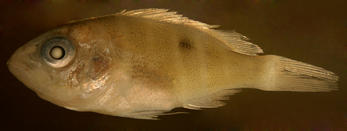
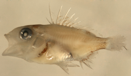
| Lutjanus
(griseus) larva |
| 6.0 mm SL |
note post-cleithral
spine, melanophores at
base of dorsal-fin spine membranes |
| San Blas, Panama, SB81-040 |
|
 |
| |
 |
|
| |
|
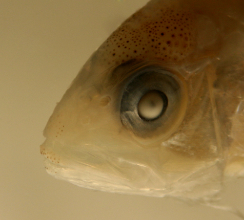
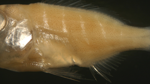
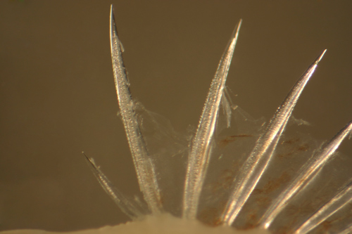
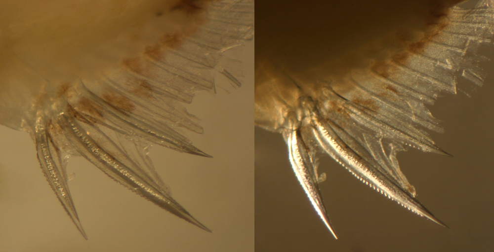
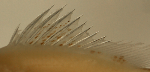
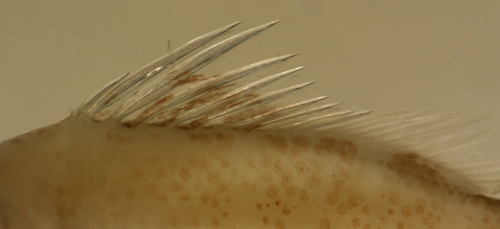
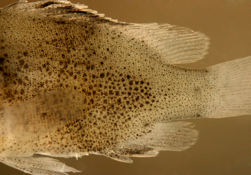
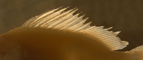
Late-Stage Larvae:
Lutjanid larvae in general share a number
of basic features, most particularly a long
non-serrated preopercular spine. The spine
decreases in length during transition and
disappears in juveniles. There are also smaller
spines lining the lower and posterior margins
of the preopercle that similarly decrease
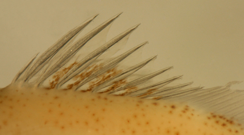
in prominence during transition. Some pretransitional
larvae can show a row of fine serrations along
the supraorbital bony ridge (preopercular
spine and supraorbital serrations visible
at the top of the photograph below of a 12.2
mm SL Gray Snapper larva, L.
griseus). |
|
 |
| |
|
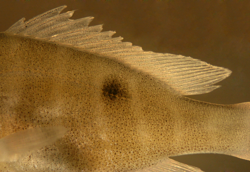
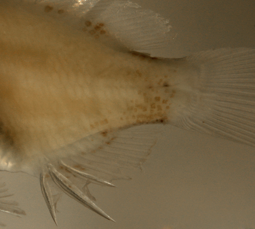
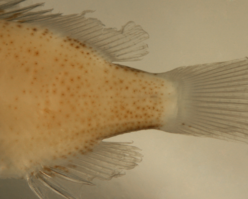
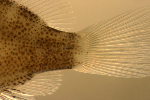
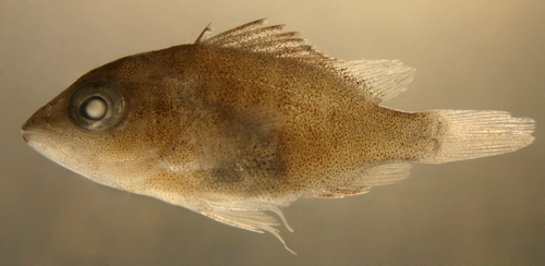
Marking patterns
on the late larvae of most snappers can be
quite similar, and comprise variations of
the basic theme of mostly dorsal-facing melanophores.
This suggests that melanophores function as
shielding, protecting vulnerable organs from
sunlight. Indeed, it would be plausible to
infer from this pattern that snapper larvae
are living near the ocean surface during the
day. Melanophores shield the brain and spinal
column by running along the top of the brain
itself, at the surface over the braincase,
along the dorsal fins, and along the dorsal
caudal peduncle. Internal melanophores line
the dorsal aspect of the vertebral column,
often with an additional short row beside
or below the column near the tail. Melanophores
line the dorsal surface of the swim-bladder
and peritoneum (overlying the abdominal organs).
In addition, the inner-facing cleithrum (the
lower rear wall of the gill cavity) is pigmented
and overlies thoracic structures. Additional
melanophores present on most species' larvae
include a few deep at the lateral midline
on the caudal peduncle, a ventral midline
caudal peduncle row (often just one or two
large melanophores), a few at the insertion
of the lower caudal-fin segmented rays, and
along the base of the membranes of the anal-fin
rays. A distinctive deep melanophore is present
from the early stages under the pterygiophores
of the last anal-fin rays (an additional "repeat"
melanophore is sometimes present on the next
segment anteriorly). These internal melanophores
can be seen on the transitional larval Yellowtail
Snapper photograph below (Ocyurus
chrysurus, 17.0 mm SL) and beginning
in the early larva of L.
griseus, photographed above). |
|

|
|
|
|
|
|
|
| |
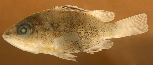
Ocyurus chrysurus
(Lutjanus chrysurus) |
|
|
|
|
| Diagnosis:
Modal fin-ray counts of D-X,13 A-III,9 indicate
Ocyurus chrysurus.
(DNA) |
| |
| Description:
Body wide and relatively thick with a sloping forehead
and a large round eye and large terminal mouth.
Dorsal-fin base long and anal-fin base short. Prominent
dorsal, anal, and pelvic-fin spines and a large
non-serrated preopercular spine. |
|
Pretransitional
mostly unmarked stage, usually from 12-17 mm SL:
Body: Pretransitional larvae develop a row
of melanophores on the side of the body near the
base of the dorsal fin. The row starts as a series
of short angled lines along the anterior aspect
of each pterygiophore below the soft dorsal fin,
then small melanophores fill in the row. The row
extends forward on the body below the spinous portion
of the fin, first as a few spots beneath the seventh
and eighth spines and the ninth and tenth spines,
and then filling in, up to the level of the third
dorsal-fin spine. On the dorsal midline of the caudal
peduncle the two lateral rows merge into a single
band of melanophores extending to the start of the
procurrent caudal-fin rays. A similar band develops
along the ventral midline of the caudal peduncle
extending forward and ending just before a single
large melanophore underlying the pterygiophores
of the last anal-fin rays. There are a few deep
melanophores at the end of the lateral midline of
the caudal peduncle and a fine speckling of small
surface melanophores around the central caudal peduncle
extending forward in a thin line along the lateral
midline. There is a single large melanophore underlying
the pterygiophores of the last anal-fin rays. A
series of short angled lines of small melanophores
develops along the anterior aspect of the anal-fin
pterygiophores, starting between the second and
fifth fin rays.
Head: Melanophores on the head consist of
dense patches overlying the brain and on the surface
braincase. There are small melanophores around the
tip of the upper jaw, along the adjacent snout,
and along the tip of the lower jaw. The opercular
area is covered in iridescence extending down to
the pelvic-fin insertion. The inner cleithral surface
of the gill cavity is speckled with large melanophores
and there are internal melanophores lining the dorsal
aspect of the swim bladder and peritoneum extending
down to the vent and overlain by a silvery camouflage
layer.
Fin Spines: The dorsal and anal-fin spines
are relatively slender, without prominent internal
reticulations. The second to fifth dorsal-fin spines
are about the same length (the second sometimes
shorter). The anal-fin spines do not show anterior
serrations. The third anal-fin spine is notably
usually longer than the second (the tip of the third
almost always extending farther back than the tip
of the second when folded down).
Fins: Melanophores are present along most
of the length of the membrane just behind the second
dorsal-fin spine and then near the outer edges of
most of the subsequent membranes of the spinous
portion of the dorsal fin. There are a few melanophores
between the bases of the lower central caudal-fin
segmented rays and the occasional individual has,
at most, one or two melanophores at the base of
the lowest of the upper caudal-fin segmented rays.
A row of melanophores develops along the anal-fin
base, one at the base of each anal-fin-ray membrane,
often including the membrane behind the third-spine.
Some individuals have melanophores on the distal
half of the two longest pelvic-fin rays.
Pretransitional analogues:
Pretransitional larvae (mostly-unmarked
stage, usually from 12-17 mm SL) can be separated
from the other regional snappers by the dorsal-fin-ray
count. In addition, the third anal-fin spine is
about the same size as the second, unlike the
Lutjanus species where the second anal-fin
spine is distinctly stouter and usually longer.
Additional useful distinguishing features include
the dorsal and anal-fin spines relatively slender
(shared with L.
cyanopterus and L.
analis), the second to fifth dorsal-fin
spines about the same length, the anal-fin spines
without prominent anterior serrations (vs. L.
griseus, L.
apodus and L.
jocu), no lateral spot or bars, and a
thin stripe of small surface melanophores extending
forward along the lateral midline from the center
of the caudal peduncle. The occasional individual
has at most one or two melanophores at the base
of the lowest of the upper caudal-fin segmented
rays (vs. several in L.
synagris and L.
mahogoni).
|
|
|
Transitional stage:
Transitional O. chrysurus larvae develop
a mostly-uniform scattering of small melanophores
on the body. Early in transition, a line of fine
surface melanophores extends forward from the
caudal peduncle along the lateral midline.
Transitional analogues:
In addition to the fin-ray counts and the similar
second and third anal-fin spines, transitional
O. chrysurus larvae can be distinguished
by the absence of spots and bars and the development
of the characteristic thin line of small melanophores
along the lateral midline.
|
|
|
Juveniles:
Juvenile O. chrysurus develop a pale midline
lateral stripe (yellow in live specimens).
Juvenile analogues:
The absence of a lateral spot or bars and a yellow
midline stripe is diagnostic.
|
|
|
|
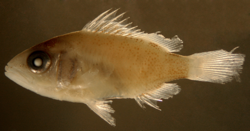
| Ocyurus
chrysurus larva |
| 15.0 mm SL |
| San Blas, Panama, SB81-000 |
|
 |
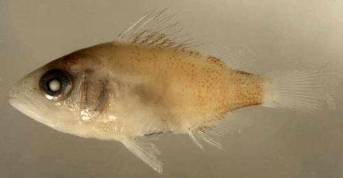
| Ocyurus
chrysurus larva |
| 16.7 mm SL |
| San Blas, Panama, SB81-024 |
|
 |
|
|
|
|
|
| Identifying transitional
Lutjanus |
|
|
|
|
|
|
|
|
|
Distinguishing the larvae and juveniles of the
numerous Lutjanus species in the region
can be difficult since many share the basic body
form as well as most fin-ray counts. Fortunately,
two common species, both with a lateral spot,
do separate out by meristics:
L. mahogoni and
L. synagris have only twelve vs. the
typical 14 dorsal-fin soft rays for the genus.
Beyond this, distinctions can be difficult since
pre-transitional larvae often have few identifying
markings. It is likely that many pre-transitional
snapper larvae will require molecular identification,
with equipment
leasing for DNA sequencing for species identification.
Transitional and juvenile snappers can also share
many of the basic markings that later distinguish
the species (such as lateral spots, incipient
bar patterns, and eye stripes). This pattern of
earlier stages sharing characters that later diverge
is commonly seen among reef fishes.
The spot snappers
The three shallow-water spot
snappers (the Lane Snapper L.
synagris, Mahogany Snapper L.
mahogoni, and Mutton Snapper L.
analis) are easily confused as larvae
and juveniles. Unlike most fishes, these snappers
converge even more in appearance after they settle
than in the transitional stages. Notably, the
relative dorsal-fin spine lengths and various
spot and bar configurations that separate species
well at transition can overlap to some degree
as small juveniles. Subtle color-pattern differences
are key to separating the larger juveniles. The
series below shows transitional recruits captured
on their first few days on the reef, when they
can still be relatively easily distinguished.


The barred snappers
There is a great deal of individual variation
in the marking patterns of transitional larvae
and recruits of the barred snappers (the Gray
Snapper L.
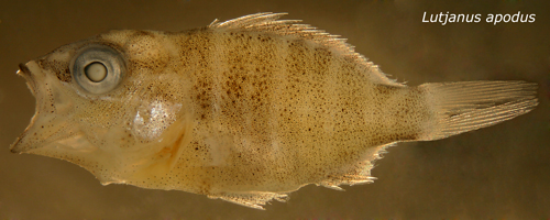
griseus, Schoolmaster Snapper
L. apodus, and Dog Snapper L.
jocu). These snappers can all display
stripes and/or bars or uniform speckling to some
degree immediately after settlement and only cleanly
diverge a week or two after settlement. For example,
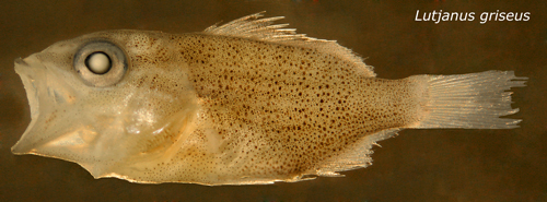
immediately after settlement some Gray Snappers
can show the vertical bars characteristic of Schoolmaster
Snappers. However, on Gray Snappers the bars tend
to fade near the anal fin (see photo below). Similarly,
some Gray Snappers are uniformly speckled before
they develop their characteristic striping and
thus look similar to newly recruited Dog Snappers,
however the latter typically have finer speckles.
Some individuals can appear intermediate and would
require DNA sequencing. Nevertheless, the vast
majority of newly-settled snappers, even those
of this difficult clade, can be identified to
species using the characters discussed below.
|
| |
|
The deep snappers
DNA sequence matching on my specimens has clarified
the identification of the late-stage larvae of
the shallow-water snappers of the region. The
deeper-water species, L.
buccanella, L.
campechanus, and L.
vivanus, however, await more comprehensive
sampling for a similarly complete treatment.
|
|
|
|
|
|
|
|
|
|
| |
|
|
|
|
|
| Diagnosis:
Modal fin-ray counts of D-X,12 A-III,8 indicate
Lutjanus synagris or L.
mahogoni. Juveniles with the lateral line
running through the lower third of the lateral spot
indicate L. synagris. (DNA) |
| |
| Description:
Body wide and relatively thick with a sloping forehead
and a large round eye and large terminal mouth.
Dorsal-fin base long and anal-fin base short. Prominent
dorsal, anal, and pelvic-fin spines and a large
non-serrated preopercular spine. |
|
|
Pretransitional mostly unmarked stage,
usually from 13-19 mm SL:
Body: Pretransitional larvae develop a
row of melanophores on the body near the base
of the dorsal fin, first along the pterygiophores
supporting the soft dorsal fin (starting between
the second and sixth rays) and then extending
below the spinous portion as a few spots between
the seventh and eighth spines and the ninth and
tenth spines. As development continues, the row
of melanophores below the dorsal fin fills in
and extends forward to the level of the third
spine (the base of the last three soft rays remain
unmarked well into transition). On the caudal
peduncle, melanophores line the dorsal and ventral
midlines and, at the lateral midline, there is
a row of deep melanophores along with a few scattered
surface melanophores. One or two discrete internal
melanophores lie well below the pterygiophores
of the last few anal-fin rays.
Head: Melanophores on the head consist
of dense patches overlying the brain and on the
surface braincase. There are small melanophores
around the tips of the upper and lower jaws. The
opercular area is covered in iridescence extending
down to the pelvic-fin insertion. The inner cleithral
surface of the gill cavity is speckled with large
melanophores and there are internal melanophores
lining the dorsal aspect of the swim bladder and
peritoneum extending down to the vent and overlain
by a silvery camouflage layer.
Fin Spines: The dorsal and anal-fin spines
are relatively stout, usually with some internal
reticulations. The anal-fin spines do not show
anterior serrations. The second dorsal-fin spine
is shorter than the third.
Fins: Small melanophores speckle the distal
two-thirds of the dorsal-fin-spine membranes.
On the caudal fin there is a vertical line of
melanophores at the base of some of the upper
as well as most of the lower segmented rays. A
row of melanophores develops along the anal-fin
base, one at the base of each soft fin ray.
Pretransitional analogues:
Pretransitional larvae of the two 12-dorsal-rayed
snappers are best distinguished by the relative
lengths of the second and third dorsal-fin spines:
in L. synagris the second dorsal-fin spine
is shorter than the third spine vs. longer (pretransitional
larvae) to about equal (late in transition) to
the third spine in L.
mahogoni. The two 12-dorsal-rayed snappers
can be separated from the 14-dorsal-rayed snappers
by the dorsal-fin-ray count, as well as having
melanophores at the bases of the upper as well
as the lower caudal-fin segmented rays at this
early stage (sometimes shared by L.
analis). The dorsal and anal-fin spines
of larval L. speciessynagris are
also stouter than in L.
analis, L.
cyanopterus, and Ocyurus
chrysurus. L. synagris larvae
at this stage also do not have distinct anterior
serrations persisting on the anal-fin spines as
do L.
griseus, L.
apodus and L.
jocu (occasional individuals do have a
few remnant serrations). Almost all pretransitional
L. synagris captured over the reef already
show the lateral spot, unlike L.
analis and the barred species.
|
|
|
Transitional stage:
Transitional L. synagris larvae develop
a prominent lateral spot early, usually with the
lateral line running through the lower third of
the spot. Iridescent bars form at each side of
the lateral spot, bracketing the black spot up
to the base of the dorsal fin. The dark bar anterior
to the spot curves away, forming a bracket. An
additional set of thin iridescent stripes develops
along the lower side alternating with thin stripes
of melanophores (in life the pale stripes are
yellow).
Transitional analogues:
The main difference between the two 12-rayed species
is that the second dorsal-fin spine is shorter
than the third in L. synagris vs. longer
or equal in L.
mahogoni (and L.
analis). The location of the lateral spot
usually differs, although some individuals do
overlap: the lateral line usually runs through
the lower third of the spot in L. synagris
and through the middle of the spot in L.
mahogoni. A consistent difference is that
on L. synagris the bar forward of the lateral
spot is not straight; it clearly curves away and
brackets the spot, while in L.
mahogoni (and L.
analis) the bar slopes evenly down from
the base of the dorsal fin across the body. Transitional
larvae of L. synagris always have a lateral
spot, then they subsequently develop bars (vs.
bars, then a spot on L.
analis). Some transitional L.
analis with a lateral spot can look remarkably
similar to L. synagris, however in the
latter the spot is large and expands the bar,
vs. staying within the bar in L.
analis. Furthermore, transitional L.
analis have three more distinct bars on
the body behind the lateral spot while these bars
are usually undeveloped on transitional L.
synagris. On new recruits, the iridescent
stripes (note, not the bars) along the lower side
of the body characteristic of L. synagris
are absent in L.
analis.
|
|
|
Juveniles:
Juvenile L. synagris have a lateral spot
with the lateral line running through the lower
third of the spot (or sometimes below the spot)
and about six to eight thick yellow stripes below
the lateral line in the bar anterior to the spot.
Juvenile analogues:
For juveniles less than 25 mm SL the relative
dorsal-spine-length characters separate juvenile
L. synagris from L.
mahogoni and L.
analis. They all have yellow stripes as
juveniles, although the stripes become thick and
more prominent on later juvenile L. synagris
and thinner and less conspicuous on juvenile L.
mahogoni. Furthermore, in juvenile L.
mahogoni the lateral spot is often
elongated (in width) and the lateral line runs
through the middle of the spot vs. a rounded or
vertically elongated spot with the lateral line
running through the lower third (or below the
spot) in L. synagris. One of the more
reliable methods to distinguish L. synagris
from L.
analis as larger juveniles is the number
of yellow stripes below the lateral line in the
bar anterior to the spot: in L.
analis the stripes tend to bifurcate into
paired thin stripes, from 9-12, while in L.
synagris the stripes remain thick and number
6-8. As they get larger, L.
analis develop a pointed outline to the
anal fin while L. synagris retains a rounded
outline.
|
|
|
|
| Lutjanus
synagris larva |
| 17.7 mm SL |
| internal melanophore
pattern |
| San Blas, Panama, SB81-039 |
|
 |
| Lutjanus
synagris |
| 16.0 mm SL |
| San Blas, Panama, SB81-000 |
|
 |
| Lutjanus
synagris transitional larva |
| 16.4 mm SL |
| note incipient bar
pattern |
|
| San Blas, Panama, SB81-047 |
|
 |
| Lutjanus
synagris transitional recruit |
| 17.8 mm SL, DNA-confirmed
ID |
| Colon, Panama, N7527b |
|
 |
| Lutjanus
synagris recruit |
| 19.3 mm SL, DNA-confirmed
ID |
| lateral spot and stripes |
| St. Thomas, USVI, ST506 |
|
 |
| |
 |
| Lutjanus
synagris recruit |
| 20.0 mm SL |
| Colon, Panama, N7527b |
|
 |
|
|
|
|
| |
|
|
|
| Diagnosis:
Modal fin-ray counts of D-X,12 A-III,8 indicate
Lutjanus mahogoni or L.
synagris. Juveniles with the lateral line
running through the middle of the lateral spot indicate
L. mahogoni. (DNA) |
|
| Description:
Body wide and relatively thick with a sloping forehead
and a large round eye and large terminal mouth.
Dorsal-fin base long and anal-fin base short. Prominent
dorsal, anal, and pelvic-fin spines and a large
non-serrated preopercular spine. |
|
|
Pretransitional mostly-unmarked stage,
usually from 17-21 mm SL:
Body: Pretransitional larvae develop a
row of melanophores on the side of the body near
the base of the dorsal fin. The row starts as
small melanophores, often lined up along the anterior
aspect of the pterygiophores below the soft dorsal
fin. The row fills in under the soft dorsal fin
and extends forward just below the spinous portion
of the fin, first as a few spots beneath the third
to fifth spines and the seventh to ninth, and
then filling in up to the level of the third dorsal-fin
spine. The two rows on each side of the dorsal
fin merge into a line of melanophores lining the
dorsal midline of the caudal peduncle (usually
made up of a short row of deeper and larger larval
melanophores overlain by a band of smaller melanophores).
A similar line develops along the ventral midline
of the caudal peduncle extending forward and ending
just before a single large melanophore underlying
the pterygiophores of the last anal-fin rays.
There are a few deep melanophores at the end of
the lateral midline of the caudal peduncle and
a fine speckling of small melanophores around
the central caudal peduncle.
Head: Melanophores on the head consist
of dense patches overlying the brain and on the
surface braincase. There are small melanophores
around the tips of the upper and lower jaws. The
opercular area is covered in iridescence extending
down to the pelvic-fin insertion. The inner cleithral
surface of the gill cavity is speckled with large
melanophores and there are internal melanophores
lining the dorsal aspect of the swim bladder and
peritoneum extending down to the vent and overlain
by a silvery camouflage layer.
Fin Spines: The dorsal and anal-fin spines
are relatively stout, usually with some internal
reticulations. The tip of the second dorsal-fin
spine often curves slightly upward (the preopercular
spine often curves slightly upward as well). The
second dorsal-fin spine is usually longer than
the third and typically the tip overlaps or extends
beyond the tip of the third. The dorsal-fin spines
then become progressively and evenly shorter such
that the profile of the spinous tips forms a straight
downward-sloping line. The anal-fin spines do
not show anterior serrations (a rare individual
has small remnant serrations). The second anal-fin
spine is longer than the third, but the tips are
closely-approximated or the third extends slightly
farther back than the tip of the second when folded
down.
Fins: Melanophores are prominent along
most of the length of the membrane just behind
the second dorsal-fin spine and are concentrated
on the membrane tag extending beyond the spine.
Smaller melanophores speckle the outer third of
all of the subsequent membranes of the spinous
portion of the dorsal fin. On the caudal fin there
are a few small melanophores at the base of some
of the upper as well as most of the lower segmented
rays. A row of melanophores develops along the
anal-fin base, one at the base of each anal-fin-ray
membrane, often including the membrane behind
the third-spine.
Pretransitional analogues:
Pretransitional larvae of the two 12-dorsal-rayed
snappers are best distinguished by the relative
lengths of the second and third dorsal-fin spines:
in L. mahogoni the second dorsal-fin spine
is longer than the third spine with the tip often
overlapping the tip of the third vs. shorter than
the third spine in L.
synagris. L. mahogoni can be separated
from most of the 14-dorsal-rayed snappers by the
dorsal-fin-ray count, as well as by having melanophores
at the bases of the upper as well as the lower
caudal-fin segmented rays at this early stage
(sometimes shared by L.
analis). The dorsal and anal-fin spines
of larval L. mahogoni are also stouter
than in L.
analis, L.
cyanopterus, and Ocyurus
chrysurus. L. mahogoni larvae at
this stage also do not have distinct anterior
serrations persisting on the anal-fin spines as
do L.
griseus, L.
apodus and L.
jocu. Almost all pretransitional L.
mahogoni captured over the reef already show
at least a few melanophores at the lateral spot,
unlike L.
analis and the barred species.
|
|
|
Transitional stage:
Transitional L.
mahogoni larvae develop a prominent lateral
spot early, usually with the lateral line running
through the middle of the spot (although some
individuals clearly have the line running through
the lower third). The spot is wider than the bar,
distinctly expanding the outline of the bar. Larval
L. mahogoni often have an upturned preopercular
spine, but this character is not consistent later.
Transitional analogues:
The main difference between the two 12-rayed species
is that the second dorsal-fin spine is longer
than the third (sometimes about equal) for L.
mahogoni (and L.
analis) vs. shorter in L.
synagris and this difference persists
in juveniles up to 25 mm SL. The location of the
lateral spot usually differs, although some individuals
do overlap: the lateral line usually runs through
the middle of the spot in L. mahogoni and
through the lower third of the spot in L.
synagris (variable in transitional L.
analis). In addition, on L.
synagris the bar forward of the lateral
spot is not straight; it clearly curves away and
brackets the spot. Transitional larvae of L.
mahogoni always have a lateral spot, then
they subsequently develop bars (vs. bars, then
a spot on L.
analis). Some transitional larvae and
early recruits of L.
analis after they develop the lateral
spot can look remarkably similar to L. mahogoni.
Other than the soft dorsal fin-ray counts, the
species can be separated by some marking differences:
the spot is larger and more elongated in L.
mahogoni, expanding the bar from which it
develops, while in L.
analis the spot is only as wide as the
bar from which it forms. Furthermore, transitional
L.
analis have three more typically distinct
bars on the body behind the lateral spot while
these bars are usually undeveloped on transitional
L. mahogoni.
|
|
|
Juveniles:
Juvenile L. mahogoni have an elongated
lateral spot with the lateral line running through
the middle of the spot.
Juvenile analogues:
Juveniles of L. mahogoni can be distinguished
by the lateral-spot location, i.e. the lateral
line through the middle of the spot in L. mahogoni
and usually through the lower third in L.
synagris and L.
analis. Notably, the lateral spot is often
elongated (in width) in juvenile L. mahogoni
vs. rounded and within the bar in L.
analis. The relative dorsal-spine-length
differences persist in juveniles up to 25 mm SL.
All have yellow stripes as juveniles, although
the stripes become thinner and less conspicuous
on juvenile L. mahogoni and thicker and
more prominent on later juvenile L.
synagris. The preopercular outline is
not diagnostic in young stages, with L.
mahogoni larvae and juveniles not showing
the notch pattern that occurs later.
|
|
|
|
|
|
| Lutjanus
mahogoni transitional larva |
| 19.7 mm SL, DNA-confirmed
ID |
| variant, lateral line
through lower third, |
| second dorsal-fin spine
longest |
| Glover's Reef, Belize,
coll. Cormac Nolan |
|
 |
|
|
 |
| Lutjanus
mahogoni recruit |
| 17.9 mm SL, |
| second dorsal-fin spine
longest |
| Colon, Panama, N7527b |
|
 |
|
|
 |
|
|
|
|
|
|
|
|
| |
|
|
|
| Diagnosis:
Modal fin-ray counts of D-X,14 A-III,8 are
shared among most of the regional Lutjanus species,
including L. analis, L.
apodus, L.
cyanopterus, L.
griseus, L.
jocu and the deep-water snappers L.
buccanella, L.
campechanus, and L.
vivanus. L.
analis is the only shallow-water snapper
with 14 dorsal-fin rays that has a lateral spot
as juveniles and adults. (DNA) |
|
| Description:
Body wide and relatively thick with a sloping forehead
and a large round eye and large terminal mouth.
Dorsal-fin base long and anal-fin base short. Prominent
stout dorsal, anal, and pelvic-fin spines and a
large non-serrated preopercular spine. |
|
|
Pretransitional mostly-unmarked stage,
usually from 13-18 mm SL:
Body: A thin line of melanophores develops
on each side just below the base of the spinous
dorsal fin, from the third to fifth and the sixth
to eighth spines (leaving an unpigmented dorsal
midline along the base of the fin). The rows continue
along the base of the soft dorsal fin on the outer
pterygiophore segments and then merge into a single
band of melanophores lining the dorsal midline
of the caudal peduncle. There are a few deep melanophores
at the end of the lateral midline on the caudal
peduncle. Along the ventral midline of the caudal
peduncle there is a single melanophore just forward
of the first procurrent caudal-fin ray, often
followed by several more toward the last anal-fin
ray.
Head: Melanophores on the head consist
of a dense patch overlying the brain and on the
surface braincase. There are small melanophores
at the tip of the upper jaw and a small patch
extends upward along the snout. The lower jaw
is mostly unmarked, with only a few small melanophores
near the tip. The opercular area is covered in
iridescence extending down to the pelvic-fin insertion.
The inner cleithral surface of the gill cavity
is speckled with large melanophores and there
are internal melanophores lining the dorsal aspect
of the peritoneum extending down to the vent and
overlain by a silvery camouflage layer.
Fin Spines: The median-fin spines are relatively
slender, without prominent internal reticulations
and lacking anterior serrations. The second dorsal-fin
spine is longer than the third and the subsequent
spines are progressively shorter. The second anal-fin
spine is only slightly longer than the third and
the tip usually does not reach farther back than
the tip of the third.
Fins: Melanophores are present along most
of the length of the membrane just behind the
second dorsal-fin spine and then near the outer
edges of most of the subsequent membranes of the
spinous portion of the dorsal fin. There are melanophores
between the bases of the lower central caudal-fin
segmented rays and often a few smaller ones between
the bases of the lowest upper caudal-fin segmented
rays. The anal fins often have no markings but
a row of melanophores usually develops during
transition (on the membranes near the base of
each soft ray). The pelvic fins develop melanophores
along the full-length of the first two soft rays
and membranes.
Pretransitional analogues:
Pretransitional larvae (mostly-unmarked, usually
from 13-18 mm SL) have relatively slender and
smooth dorsal and anal-fin spines, without the
prominent internal reticulations and anterior
serrations found in L.
griseus, L.
apodus, and L.
jocu. In addition, amongst the regional
Lutjanus, only L. analis and L.
cyanopterus often have no melanophores
along the base of the anal-fin rays before transition.
Pretransitional L. analis also share the
relatively slender and smooth spines (and snout
melanophores) with L.
cyanopterus, but can be distinguished
by having a distinctly-narrower caudal peduncle,
melanophores along the full-length of the pelvic-fin
membranes or absent (vs. on the outer third of
the longest pelvic-fin membranes), and the tip
of the second anal-fin spine is near the tip of
the third (vs. the tip of the second usually extending
well past the tip of the third in L.
cyanopterus).
|
|
|
Transitional stage:
Transitional L.
analis larvae develop distinct bars of
small melanophores slanted down and to the rear.
The space between the seven bars is typically
iridescent and usually not speckled during transitional
stages. A lateral spot then develops in the fourth
bar at the level of the lateral line, intensifying
within the confines of the bar of melanophores
(without expanding the outline of the bar).
Transitional analogues:
In contrast to transitional L.
analis, the bars are vertical in L.
apodus,
L. jocu, and
L. cyanopterus; the bar originating
at the mid-soft-dorsal fin slants down to meet
the last anal-fin ray in L. analis vs.
to the mid-anal fin in the vertically-barred species.
Transitional L. analis often do not develop
the characteristic lateral spot until past the
transitional stage (vs. early and isolated spot
appearance in the 12-rayed
snappers). When the spot develops, it can
be difficult to separate L. analis from
L.
synagris and L.
mahogoni. A reliable difference is in
the relative length of the second dorsal-fin spine:
well shorter than the third in L.
synagris vs. longer or equal in transitional
L. analis and L.
mahogoni. The marking patterns do diverge
as the spot develops: on L. analis recruits
the spot notably forms within the bar, without
expanding the outline of the bar as it does on
the 12-rayed snappers. In addition, on L.
synagris the bar forward of the lateral
spot is not straight; it clearly curves away and
brackets the spot. Transitional L. analis
also have three typically distinct bars on the
body behind the one with the lateral spot while
these bars are usually indistinct on transitional
larvae of the others.
|
|
|
Juveniles:
Juvenile L. analis have a distinct round
lateral spot contained within the confines of
a dark bar, with the lateral line usually running
through the lower third and more than eight yellow
stripes on the body below the lateral line in
the bar anterior to the spot.
Juvenile analogues:
Juvenile L. analis can be separated from
the other shallow-water species with bars and
a lateral spot by the 14 soft dorsal-fin rays
(vs. 12 in L.
synagris and L.
mahogoni). Larger juveniles of L. analis
converge in appearance with the other spotted
snappers, often sharing the lateral spot location
(spot size and location can be variable), the
striping on the side of the body, and even the
relative dorsal-fin spine lengths (after 25 mm
SL). One of the more reliable methods to distinguish
the species as larger juveniles is the number
of yellow stripes below the lateral line in the
bar anterior to the spot: in L. analis
the stripes tend to bifurcate into numerous thin
stripes, from 9-12, while in L.
synagris the stripes remain thick and
number 6-8. L.
mahogoni juveniles have inconsistent yellow
stripes, but their lateral spot is typically larger
and elongated in width compared to that of L.
analis. As they get larger L. analis
develop a pointed outline to the anal fin while
the other species retain a rounded outline.
|
|
|
|
| Lutjanus
analis larva |
| 16.0 mm SL, DNA-confirmed
ID |
|
| San Blas, Panama, SB83-169 |
|
 |
| |
 |
| |
 |
| Lutjanus
analis transitional larva |
| 16.9 mm SL |
| note incipient lateral
spot within bar |
| San Blas, Panama, SB81-053 |
|
 |
|
|
 |
| |
 |
| |
 |
| |
 |
| Lutjanus
analis transitional larva |
| 16.6 mm SL, DNA-confirmed
ID |
| slanted bars with no
lateral spot |
| Glover's Reef, Belize,
coll. Cormac Nolan |
|
 |
| Lutjanus
analis transitional recruit |
| 15.4 mm SL, DNA-confirmed
ID |
| Colon, Panama N7527b |
|
 |
| Lutjanus
analis recruit |
| 17.6 mm SL, DNA-confirmed
ID |
| Colon, Panama N7527b |
|
 |
| Lutjanus
analis recruit |
| 23.2 mm SL, DNA-confirmed
ID |
| Colon, Panama N7527b |
|
 |
| Lutjanus
analis juvenile |
| about 30 mm SL |
| spot within bar, lateral
line in lower third, |
| more than 8 yellow
stripes below lat line |
| courtesy Garold Sneegas |
|
 |
| |
|
|
|
| |
|
|
| |
|
|
|
| Diagnosis:
Modal fin-ray counts of D-X,14 A-III,8 are
shared among most of the regional Lutjanus species,
including L.
analis, L.
apodus, L.
cyanopterus, L. griseus, L.
jocu and the deep-water snappers L.
buccanella, L.
campechanus, and L.
vivanus. Juvenile L.
griseus have a prominent dark stripe through
the eye, a dark striped body and no lateral spot.
(DNA) |
|
| Description:
Body wide and relatively thick with a sloping forehead
and a large round eye and large terminal mouth.
Dorsal-fin base long and anal-fin base short. Prominent
dorsal, anal, and pelvic-fin spines and a large
non-serrated preopercular spine. |
|
|
Pretransitional mostly-unmarked stage,
usually from 10-13 mm SL:
Body: Pretransitional larvae have two patches
of melanophores on the body below the dorsal fin:
under the last two dorsal-fin spines and first
dorsal-fin soft rays and then under the middle
of the soft dorsal fin. There is a band of surface
melanophores along the anterior half of the dorsal
midline of the caudal peduncle and full-length
along the ventral midline of the caudal peduncle,
extending forward and ending just before a single
large melanophore underlying the pterygiophores
of the last anal-fin rays. A patch of surface
melanophores develops on the caudal peduncle filling
in progressively from ventral to dorsal in a mostly
uniform pattern, without a distinct clear bar
anteriorly on the lower half of the caudal peduncle.
There are a few deep melanophores at the end of
the lateral midline of the caudal peduncle.
Head: Melanophores on the head consist
of a patch overlying the brain and on the surface
braincase and around the tip of the upper jaw;
at the tip of the lower jaw there are either no
melanophores or distinctly fewer than at the tip
of the upper jaw. The opercular area is covered
in iridescence extending down to the pelvic-fin
insertion. The inner cleithral surface of the
gill cavity is speckled with large melanophores
and there are internal melanophores lining the
dorsal aspect of the peritoneum extending down
to the vent and overlain by a silvery camouflage
layer.
Fin Spines: The dorsal and anal-fin spines
are relatively stout, with prominent internal
reticulations. There are fine serrations along
the anterior aspect of the anal and dorsal-fin
spines at this stage, typically persisting into
transition in this species.
Fins: Melanophores on the dorsal-fin membranes
are concentrated between the third and eighth
spines, present on the dorsal midline at the base
of the membrane and extending halfway or two-thirds
up the membranes. The dorsal-midline melanophores
are often present from the second to the tenth
dorsal-fin spine bases. On the anal fin, there
are melanophores on the lower half of the membranes
between the anal-fin spines and about half-way
up the second anal-fin spine. They continue on
the lower half of the membrane between the last
spine and the first ray and the next membrane,
followed by melanophores only at the base of the
membrane for the next few rays. Melanophores are
concentrated below the pterygiophores of the last
two or three rays (often as one conspicuous large
melanophore) where they join the row along the
ventral midline of the caudal peduncle. There
are often a few melanophores along the proximal
portion of the segmented caudal-fin rays in two
places: between the bases of the lower-central
rays and along the lowest two or three rays, the
latter often extending in a line out along the
rays.
Pretransitional analogues:
Pretransitional larvae (mostly-unmarked stage,
usually from 10-13 mm SL) are separated from many
other Lutjanus by having distinct serrations
persisting on the anterior profile of the anal
and dorsal-fin spines (but shared by L.
apodus and L.
jocu). The L. griseus larval type
is distinguished from the L.
apodus and L.
jocu types at this stage by having the
spinous-dorsal-fin melanophores mostly on the
proximal two-thirds of the membranes with some
touching the dorsal midline around the insertion
of the spines (vs. melanophores concentrated on
the distal portion of the membranes and sparing
the dorsal midline at the base of most of the
dorsal-fin spines). Additional features separating
lightly marked L. griseus from L.
apodus are a more uniform melanophore
scattering on the lower caudal peduncle (vs. concentrating
as a bar at the posterior half), melanophores
along the anterior half of the dorsal midline
of the caudal peduncle (vs. a short line usually
less than a third of the peduncle length), more
melanophores on the tip of the upper than lower
jaws, and relatively lightly or unmarked lower
head and pelvic fins (even on heavier-marked larvae).
|
|
|
Transitional stage:
Transitional larvae of L. griseus develop
a relatively uniform scattering of melanophores
on the body, usually with some indistinct light
bars on the upper side of the body. The lower
half of the caudal peduncle is uniformly speckled.
Melanophores extend from the dorsal midline out
to about two-thirds of the spinous-dorsal-fin
membranes. A distinct stripe extends from the
eye forward to the tip of the upper jaw and two
stripes develop behind the eye and diverge. The
tip of the lower jaw, the ventral half of the
head and the pelvic fins are lightly speckled
in most transitional larvae. Transitional recruits
are mostly uniformly speckled; when indistinct
bars are present they fade out on the lower body,
especially near the anal fin. Many show a pattern
of large blotchy melanophores over a finer speckling.
The large melanophores disappear in small juveniles
and are replaced by a pattern of stripes.
Transitional analogues:
Transitional L. griseus larvae tend
to have a relatively uniform scattering of melanophores
on the body with no lateral spot, distinguishing
them from the other spotted or barred species.
Furthermore, transitional L. griseus larvae
usually retain anterior serrations on the anal-fin
spines separating them from most other snappers
(except some L.
apodus and L.
jocu). Some transitional L. griseus
may have indistinct bars, but these are typically
limited to the upper half of the body vs. obvious
full-body bars in larval L.
apodus and L.
analis. Especially on early-transitional
stages, L. griseus have a lightly-marked
lower jaw and head below the level of the eye
(vs. often heavily spotted in the latter species),
and pelvic fins mostly unpigmented (vs. covered
in melanophores). The other uniformly-marked transitional
snappers comprise L.
jocu and L.
cyanopterus. Transitional L.
cyanopterus have a quite different body
shape with a narrower and longer body and a wider
caudal peduncle (relative to body depth). Transitional
recruits of L.
jocu can appear quite similar to transitional
L. griseus, but in L.
jocu the thin indistinct light bars persist
without any development of body stripes and the
body is more finely speckled.
|
|
|
Juveniles:
Juvenile L. griseus are overall dusky with
a prominent dark stripe through the eye and a
pattern of thin parallel dark lines across the
body, most distinctly below the lateral line.
The stripes are characteristically made up of
rows of individual dark-spotted scales. Some individuals
show an indistinct bar pattern, but it is limited
to the upper half of the body.
Juvenile analogues:
L. griseus juveniles lack the lateral spot
of many other snapper juveniles and do not show
the prominent bars of juvenile L.
apodus. They are wider-bodied with a narrower
caudal peduncle than juvenile L.
cyanopterus. L. griseus juveniles
can appear similar to some of the more uniformly-marked
early juvenile L.
apodus and L.
jocu, however the thin dark stripes on
the side of the body and the dark stripe through
the eye intensify in L. griseus, while
in juvenile L.
jocu the dark stripe through the eye becomes
less prominent and the blue line below the eye
intensifies and juvenile L.
apodus rapidly develop prominent bars.
|
|
|
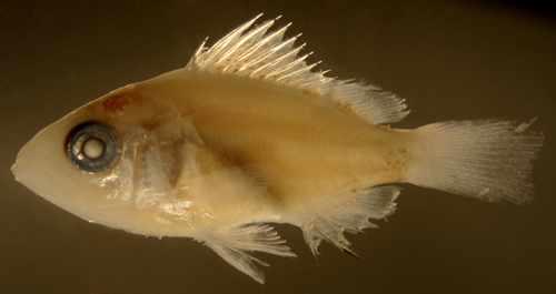
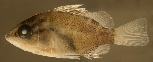
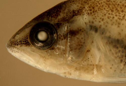
| Lutjanus
griseus larva |
| 12.2 mm SL |
| anterior spine serrations/cleithral
pigment |
| San Blas, Panama, SB81-019 |
|
 |
| |
 |
| |
 |
 |
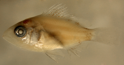
| Lutjanus
griseus larva |
| 12.6 mm SL. DNA-confirmed
ID |
| |
| San Blas, Panama, SB81-059 |
|
 |
| |
 |
| |
 |
| |
 |
| Lutjanus
griseus early transitional larva |
| 13.7 mm SL |
|
| San Blas, Panama, SB81-018B |
|
 |
| |
 |
| |
 |
| |
 |
| Lutjanus
griseus transitional larva |
| 12.4 mm SL, DNA-confirmed
ID |
| note body widening,
3rd spine enlarging |
| San Blas, Panama, SB86-701 |
|
 |
| |
 |
| |
 |
| |
 |
| Lutjanus
griseus transitional larva |
| 13.0 mm SL, DNA-confirmed
ID |
| indistinct bar pattern |
| Glover's Reef, Belize,
coll. Cormac Nolan |
|
 |
| |
 |
| |
 |
| Lutjanus
griseus transitional recruit |
| 12.9 mm SL, DNA ID
confirmed |
| Isla Grande, Panama,
N7528b |
|
 |
| Lutjanus
griseus transitional recruit |
| 13.9 mm SL, DNA ID
confirmed |
| speckled transitional
variant |
| Isla Grande, Panama,
N7528b |
|
 |
| |
 |
| Lutjanus
griseus, transitional recruit |
| 15.0 mm SL, DNA ID
confirmed |
| indistinct bars more
on upper body |
Colon,
Panama, N7527b
. |
|
 |
|
 |
| Lutjanus
griseus, transitional recruit |
| 14.8 mm SL, DNA ID
confirmed |
| speckled, incipient
stripes |
| Isla Grande, Panama,
N7528b |
|
 |
| |
 |
| Lutjanus
griseus recruit |
| 18.0 mm SL, DNA ID
confirmed |
| Isla Grande, Panama,
N7528b |
|
 |
|
|
|
|
| |
|
|
|
| Diagnosis:
Modal fin-ray counts of D-X,14 A-III,8 are
shared among most of the regional Lutjanus species,
including L.
analis, L. apodus, L.
cyanopterus, L.
griseus, L.
jocu and the deep-water snappers L.
buccanella, L.
campechanus, and L.
vivanus. Transitional and juvenile L.
apodus have a prominent pattern of vertical
bars without a lateral spot. (DNA) |
|
| Description:
Body wide and relatively thick with a sloping forehead
and a large round eye and large terminal mouth.
Dorsal-fin base long and anal-fin base short. Prominent
dorsal, anal, and pelvic-fin spines and a large
non-serrated preopercular spine. |
|
|
Pretransitional mostly-unmarked stage,
usually from 10-15 mm SL:
Body: Pretransitional larvae can have a
few melanophores on the body just below the dorsal
fin base where the future bars will develop: at
the mid-spinous dorsal fin, the end of the spinous
dorsal fin and under the mid-soft dorsal fin.
There is a patch of melanophores along the anterior
third of the dorsal midline of the caudal peduncle
and a full-length band of melanophores along the
ventral midline of the caudal peduncle extending
forward and ending just before a single large
melanophore underlying the pterygiophores of the
last anal-fin rays. A bar pattern begins on the
lower caudal peduncle as a patch without melanophores
between two bars. There are a few deep melanophores
at the very end of the lateral midline of the
caudal peduncle.
Head: Melanophores on the head consist
of a patch overlying the brain and on the surface
braincase and small melanophores around the tips
of both the upper and lower jaw (in similar numbers).
The opercular area is covered in iridescence extending
down to the pelvic-fin insertion. The inner cleithral
surface of the gill cavity is speckled with large
melanophores and there are internal melanophores
lining the dorsal aspect of the peritoneum extending
down to the vent and overlain by a silvery camouflage
layer.
Fin Spines: The dorsal and anal-fin spines
are relatively stout, with prominent internal
reticulations. There are fine serrations along
the anterior aspect of the anal-fin spines at
this stage, but disappearing during transition.
Fins: Melanophores on the dorsal-fin membranes
are concentrated between the third and eighth
dorsal-fin spines, predominantly on the distal
half of the fin-ray membranes and typically sparing
the membranes adjacent to the base of the fin.
Some individuals have melanophores spreading down
to meet the dorsal midline, but only behind the
fourth and fifth spines and later the ninth and
tenth spines (at the site of the future dark bars
on the body). On the anal fin, there are melanophores
along the base of the spines and membranes, spreading
almost half-way up the second and third anal-fin
spines. There are melanophores on the lower portion
of the membrane between the last anal-fin spine
and the first ray and the next membrane or two,
followed by melanophores at the base of the membrane
for the next few rays. There can be a few melanophores
between the bases of the uppermost of the lower
segmented caudal-fin rays and also on the lowest
two or three segmented rays, often extending out
along the rays.
Pretransitional analogues:
Pretransitional larvae (mostly-unmarked stage,
usually from 10-15 mm SL) are separated from some
other Lutjanus by having distinct serrations
persisting on the anterior profile of the anal
and dorsal-fin spines (but shared by L.
griseus and L.
jocu). L. apodus larvae are distinguished
from the L.
griseus type at this stage by having the
dorsal-fin membrane melanophores concentrated
on the distal portion of the membranes, only a
small patch on the anterior third of the dorsal
midline of the caudal peduncle, usually an incipient
bar pattern on the lower caudal peduncle, similar
numbers of melanophores on the tip of the upper
and lower jaws, and often pigmentation along the
longest pelvic-fin membrane. It is likely that
pretransitional L.
jocu cannot be separated from L. apodus.
|
|
|
Transitional stage:
Early transitional
L. apodus develop a pattern of
bars on the body, beginning at the lower caudal
peduncle where two dark bars first separate and
then bars progressively develop from the caudal
peduncle anteriorly. Each bar starts below the
base of the dorsal fin and extends down with development.
The mid-body bars begin with three patches of
melanophores: the first under the fourth to sixth
dorsal-fin spines, the second under the last two
dorsal-fin spines and first dorsal soft rays and
the third under the middle of the soft dorsal
fin. Melanophores are limited to the outer half
of the spinous-dorsal-fin membranes at first,
but progressively extend down during transition.
Before any stripes develop on the head, the tips
of the upper and lower jaws are similarly speckled
with small melanophores. Even on lightly-marked
transitional larvae, there are some small melanophores
on the thorax and the pelvic fin membranes. Early
transitional larvae have serrations on the anterior
aspect of the first two anal-fin spines, but these
are usually lost midway through transition. Late
transitional larvae have melanophores covering
much of the body, but now the bars are made up
of alternating areas of smaller and larger melanophores.
The lower caudal peduncle at this stage has filled-in
with melanophores and no longer has bars separated
by non-pigmented skin. By this point, melanophores
have advanced down the spinous-dorsal-fin membranes
and do reach the base where they merge with the
melanophores of the darker bars. On the head,
a stripe develops between the eye and the tip
of the upper jaw and two stripes diverge behind
the eye. Small melanophores fill in and uniformly
speckle the cheek, operculum, and thorax as well
as the pelvic fins.
Transitional analogues:
Transitional L. apodus larvae develop bars
on the body with no lateral spot, distinguishing
them from the spotted species. In the early stages
of transition, L. apodus larvae can be
separated from L.
griseus by having incipient melanophore
bars forming on the lower caudal peduncle (vs.
uniform) and having melanophores mostly on the
distal half of the spinous-dorsal-fin membranes
(vs. proximal and base). Late transitional L.
apodus larvae differ from L.
griseus by having distinct vertical bars
of larger melanophores on the body vs. uniform
speckling over the upper body (and lighter over
the lower half of the head and abdomen) or indistinct
bars at most on the upper half of the body, numerous
melanophores speckling the cheek, thorax, and
pelvic fins (vs. those areas relatively lightly-marked).
Transitional recruits of L.
jocu can overlap in appearance and can
show a similar bar pattern to that of transitional
L. apodus, although the lighter bars are
narrower and the bar pattern becomes less distinct
with development. There is also some overlap in
appearance when both have a mostly-uniform speckling
pattern, although uniform L. apodus have
large melanophores and L.
jocu have a fine speckling of melanophores
(for example, in the space below the eye, there
are about 100 melanophores in an area equal to
the pupil in L.
jocu vs. about 10 in L. apodus).
Transitional recruits of L. apodus and
L.
griseus can sometimes overlap in appearance.
If the bars are distinct from the base of the
dorsal fin down to the anal fin, it is L. apodus;
when L.
griseus have bars, they are apparent on
the upper body but fade towards the anal-fin base.
As they grow, L.
griseus develop distinct striping patterns
on the lower body that do not occur on L. apodus.
|
| |
Juveniles:
Juvenile L. apodus have prominent vertical
bars and no lateral spot. Rare individuals have
a uniform pattern with only indistinct bars, but,
notably these individuals do not show any striping
pattern.
Juvenile analogues:
The absence of a lateral spot separates L.
apodus from most other juvenile snappers ((L.
analis, L.
mahogoni,
L. synagris, and the deep-water snappers).
Virtually all juvenile L. apodus have prominent
vertical bars which are absent or indistinct in
L. jocu,
L.
griseus, and L.
cyanopterus. Rare individuals of L.
apodus that have a uniform appearance or indistinct
bars can be difficult to separate from juvenile
L. jocu,
but juvenile L.
griseus of the same size would show some
evidence of body stripes. Juvenile L.
cyanopterus are narrower-bodied, have
a wider caudal peduncle, and do not share the
blue line under the eye.
|
|
|
|
|
| Lutjanus
apodus larva |
| 14.1 mm SL |
| |
| San Blas, Panama, SB81-053 |
|
 |
| |
 |
| |
 |
| |
 |
| Lutjanus
apodus early transitional larva |
| 12.6 mm SL |
| |
| San Blas, Panama, SB81-054 |
|
 |
| |
 |
| |
 |
| |
 |
| |
 |
| Lutjanus
apodus transitional larva |
| 14.4 mm SL |
| note mostly-bare dorsal
caudal peduncle |
| San Blas, Panama, SB81-053 |
|
 |
| Lutjanus
apodus late transitional larva |
| 15.8 mm SL, DNA-confirmed
ID |
| |
| San Blas, Panama, SB81-031 |
|
 |
| |
 |
| |
 |
| |
 |
| Lutjanus
apodus late transitional larva |
| 14.8 mm SL |
| distal pigmentation
on dorsal membranes |
| |
| San Blas, Panama, SB81-031 |
|
 |
| Lutjanus
apodus late transitional larva |
| 14.0 mm SL |
| variant, indistinct
bars |
| |
| San Blas, Panama, SB81-100 |
|
 |
| Lutjanus
apodus late transitional larva |
| 16.6 mm SL, DNA-confirmed
ID |
| indistinct bars |
| |
| Barbados, V05-820,
coll. by Henri Valles |
|
 |
| Lutjanus
apodus transitional recruit |
| 13.8 mm SL, DNA-confirmed
ID |
|
| |
| Glover's reef, Belize,
coll. Cormac Nolan |
|
 |
| Lutjanus
apodus transitional recruit |
| 13.5 mm SL, DNA-confirmed
ID |
| bars indistinct, but
extend to lower body |
|
| |
Colon, Panama, N7527b
. |
|
|
|
 |
| Lutjanus
apodus transitional recruit |
| 18.5 mm SL, DNA-confirmed
ID |
| bars indistinct, but
extend to lower body |
|
| |
Colon, Panama, N7527b
. |
|
|
|
 |
| |
|
|
|
| |
|
|
|
|
|
|
| Diagnosis:
Modal fin-ray counts of D-X,14 A-III,8 are
shared among most of the regional Lutjanus species,
including L.
analis, L.
apodus, L.
cyanopterus, L.
griseus, L. jocu and the deep-water
snappers L.
buccanella, L.
campechanus, and L.
vivanus. Juvenile L. jocu have indistinct
vertical bars, no lateral spot, and a prominent
blue line from under the eye to the maxilla. Juveniles
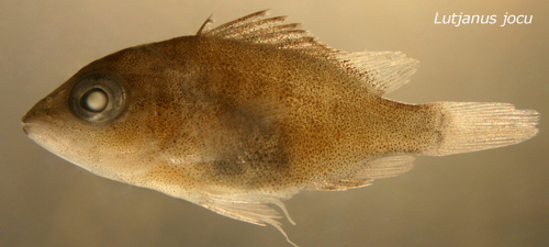
are found in mangrove habitats. (DNA) |
|
| Description:
Body wide and relatively thick with a sloping forehead
and a large round eye and large terminal mouth.
Dorsal-fin base long and anal-fin base short. Prominent
dorsal, anal, and pelvic-fin spines and a large
non-serrated preopercular spine. |
|
|
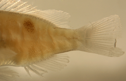
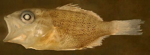
Transitional stage:
Transitional recruits of L. jocu have a
mostly-uniform scattering of fine melanophores
on the body with notably indistinct bars against
a finely-speckled background. The blue stripe
from under the eye to the mid-maxilla is prominent.
Transitional analogues:
Transitional recruits of L. jocu develop
indistinct bars in the same pattern as the prominent
bars in L.
apodus. The early recruits of the two
species can be difficult to separate, but the
bars on L. jocu are generally indistinct
(particularly below the anterior dorsal-fin spines)
and absent on the caudal peduncle. An additional
difference is that the melanophores on L. jocu
begin as a very fine and dense scattering vs.
larger and sparser melanophores on transitional
L.
apodus (for example, in the space below
the eye, there are about 100 melanophores in an
area equal to the pupil in L. jocu vs.
about 10 in L.
apodus). L. jocu recruits can also
be difficult to separate from transitional L.
griseus, however the latter have large blotchy
melanophores over a fine spotted background, vs.
uniform fine speckling seen in L. jocu.
L.
griseus recruits also rapidly acquire
their characteristic striping. L.
cyanopterus early recruits share the indistinct
bars but do not have the obvious blue stripe on
the head and they retain their characteristic
black edging to the pelvic fins through the transitional
phase.
|
|
|
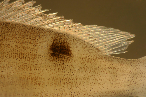
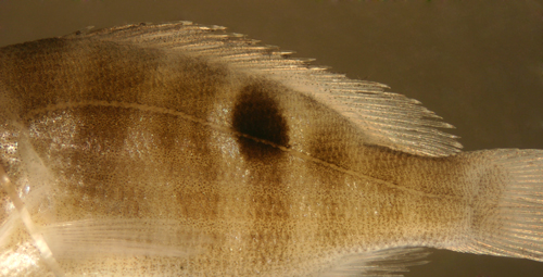
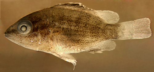
Juveniles:
Juvenile L. jocu have few distinct markings
other than the thin blue line extending from the
maxilla back under the eye and across the operculum.
Most juveniles retain some evidence of indistinct
bars.
Juvenile analogues:
Juvenile L. jocu have no lateral spot (vs.
L.
analis, L.
mahogoni,
L. synagris, and the deep-water snappers)
and indistinct vertical bars (vs. prominent in
L.
apodus). They are wider-bodied than L.
cyanopterus (which lacks the blue line
under the eye as well). Juvenile L.
griseus intensify the dark stripe through
the eye and develop thin dark stripes on the side
of the body.
|
|
|
| |
| Lutjanus
jocu |
|
| 14.0 mm SL, |
| San Blas, Panama,
SB81-112 |
|
 |
| |
 |
| |
 |
| |
 |
| |
 |
|
| Lutjanus
jocu, transitional recruit |
| 15.0 mm SL, DNA-confirmed
ID |
| bars indistinct, under-eye
stripe |
| Portobelo, Panama,
n762c |
|
 |
|
|
 |
| Lutjanus
jocu, transitional recruit |
| 16.8 mm SL, DNA-confirmed
ID |
| bars indistinct, under-eye
stripe |
| Carrie Bow Cay, Belize,
1986 |
|
 |
| Lutjanus
jocu, juvenile |
| 22.9 mm SL, DNA-confirmed
ID |
indistinct vertical
bars
blue under-eye stripe |
| Isla Grande, Panama,
N7529a |
|
 |
| |
 |
| |
 |
| |
|
|
|
|
|
|
| |
|
|
|
|
An earlier version of the following description
and some of the photographs have previously been
published in Zootaxa (copyright reserved by Magnolia
Press):
Victor, B.C., Hanner, R., Shivji, M., Hyde,
J. & Caldow, C. (2009) Identification of the
larval and juvenile stages of the Cubera Snapper,
Lutjanus cyanopterus, using DNA barcoding.
Zootaxa, 2215, 24-36.
Diagnosis: Modal
fin-ray counts of D-X,14 A-III,8 are shared among
most of the regional Lutjanus, including
L. analis,
L. apodus,
L. cyanopterus, L.
griseus, L.
jocu and the deep-water snappers L.
buccanella, L.
campechanus, and L.
vivanus. Juvenile L. cyanopterus
have an indistinct barred pattern without a lateral
spot. Adult Cubera snappers are the largest Western
Atlantic snappers and can reach four feet in length
and weigh up to 125 pounds. (DNA) |
|
| Description:
Body wide and relatively thick with a sloping forehead
and a large round eye and large terminal mouth.
Dorsal-fin base long and anal-fin base short. Prominent
stout dorsal, anal, and pelvic-fin spines and a
large non-serrated preopercular spine. |
|
|
Pretransitional mostly-unmarked stage,
usually from 15-18 mm SL:
Body: A thin line of melanophores develops
on each side just below the base of the spinous
dorsal fin, from the third to sixth and then from
the eighth to tenth spines (leaving an unpigmented
dorsal midline along the base of the fin). Continuing
along the base of the soft dorsal fin, the melanophore
row widens to cover the outer pterygiophore segments,
intensifying beneath the fourth to eighth and
the last two rays and then extending along the
dorsal midline of the caudal peduncle, ending
at the start of the procurrent caudal-fin rays.
Early transitional larvae begin to develop short
melanophore streaks lining some of the myomere
edges along the mid-upper body. A central patch
of surface melanophores develops on the end of
the caudal peduncle, then filling in progressively
from ventral to dorsal. There are a few deep melanophores
at the end of the lateral midline of the caudal
peduncle. Along the ventral midline of the caudal
peduncle there is a line of melanophores starting
just after the melanophore underlying the base
of the last anal-fin rays and extending up to
the procurrent caudal-fin rays.
Head: Melanophores on the head consist
of a dense patch overlying the brain and on the
surface braincase with a scattering developing
between the braincase and the first dorsal-fin
spine. A patch of small melanophores develops
at the tip of the upper jaw and then extends upward
along the snout. The lower jaw is mostly unmarked,
with only a few small melanophores near the tip.
The opercular area is covered in iridescence extending
down to the pelvic-fin insertion. The inner cleithral
surface of the gill cavity is speckled with large
melanophores and there are internal melanophores
lining the dorsal aspect of the peritoneum extending
down to the vent and overlain by a silvery camouflage
layer.
Fin Spines: The dorsal and anal-fin spines
are relatively slender, without prominent internal
reticulations or serrations. The second dorsal-fin
spine is the longest, with the spines becoming
progressively and evenly shorter such that the
profile of the spinous tips forms a straight downward-sloping
line.
Fins: Melanophores on the dorsal-fin membranes
are present along the full length of the membrane
just behind the second dorsal-fin spine and then
densely on the outer third of all of the subsequent
spinous-dorsal-fin membranes. The soft dorsal
fin is unmarked. There are a few melanophores
between the bases of the lower central caudal-fin
segmented rays. The anal fin is unmarked. The
pelvic fins have dense melanophores along the
outer third of the fin membranes of the longest
two or three rays.
Pretransitional analogues:
Pretransitional larvae (mostly-unmarked, usually
from 15-18 mm SL) have relatively slender and
smooth dorsal and anal-fin spines, without the
prominent internal reticulations and anterior
serrations found in L.
griseus, L.
apodus, and L.
jocu. In addition, amongst the regional
Lutjanus only L. cyanopterus and
L.
analis have a mostly unmarked anal fin,
with no melanophores on the membranes or even
at teh base of most of the anal-fin elements before
transition. Pretransitional L. cyanopterus
share the relatively slender and smooth dorsal-fin
spines and snout melanophores with L.
analis, but have a distinctly-wider caudal
peduncle and melanophores on the outer third of
the longest pelvic-fin membranes (vs. full-length
or none). On larval L. cyanopterus the
dorsal-fin spines (after the first) are evenly
and progressively shorter, unlike some other species.
The anterior snout and upper jaw are speckled
with melanophores with only a few on the lower
jaw (vs. roughly similar on the upper and lower
jaw and/or not on the snout as well in other species).
L. cyanopterus larvae have a wider caudal
peduncle than other regional Lutjanus species
(body depth after last dorsal ray goes fewer than
2.5 times into body depth at first dorsal-fin
spine).
|
|
|
Juveniles:
Juvenile L. cyanopterus have few distinguishing
markings, primarily indistinct vertical bars and
a dark outer portion of the spinous dorsal fin
with abruptly-light edging. The black markings
on the outer third of the pelvic-fin membranes
shown by larvae can also persist well into the
juvenile stage. Dark variants develop a black
cap across the eyeball, uniformly-darkened pelvic-fin
membranes, and intensified dark bars on the body
and black edging to the spinous dorsal fin. Even
in the juvenile stage, Cubera snappers have markedly-enlarged
canine teeth. The body shape of juveniles and
adults differs from other regional snappers in
being longer and narrower; the body depth (predorsal)
of L. cyanopterus juveniles goes at least
2.8 times into SL (vs. 2.4 or fewer).
Juvenile analogues:
Juvenile L. cyanopterus have no lateral
spot (vs. L.
analis, L.
mahogoni,
L. synagris, and the deep-water snappers)
and lack the prominent eye stripes or blue lines
across the cheek characteristic of L.
apodus, L.
jocu and L.
griseus. In addition, the other regional
Lutjanus have distinctly wider bodies as
both juveniles and adults: the predorsal body
depth of L. cyanopterus juveniles goes
at least 2.8 times into SL (vs. 2.4 or fewer).
|
|
|
| Lutjanus
cyanopterus larva |
| 18.8 mm SL, DNA-confirmed
ID |
| San Blas, Panama, SB81-019 |
|
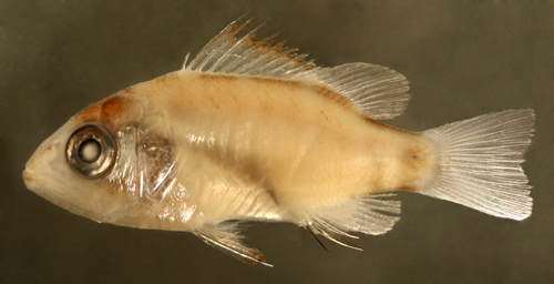 |
| |
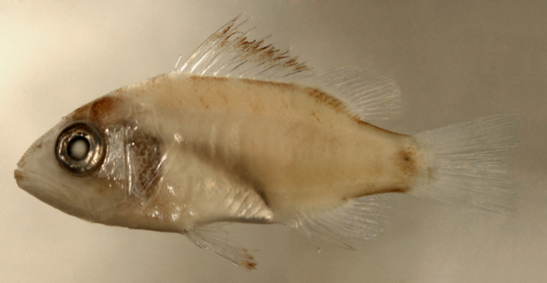 |
| |
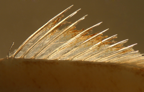 |
| |
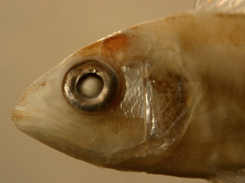 |
| Lutjanus
cyanopterus larva |
| 17.7 mm SL, DNA-confirmed
ID |
| San Blas, Panama, SB81-014 |
|
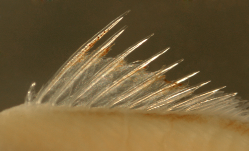 |
| Lutjanus
cyanopterus larva |
| 18.4 mm SL |
| San Blas, Panama, SB81-016 |
|
 |
| Lutjanus
cyanopterus larva |
| 18.7 mm SL |
| San Blas, Panama, SB81-015 |
|
 |
| Lutjanus
cyanopterus juvenile |
| 25.8 mm SL, DNA-confirmed
ID |
| note indistinct vertical
bars |
| pigmented outer third
of pelvic fin |
| Isla Grande, Panama,
N7529a |
|
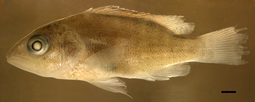 |
| |
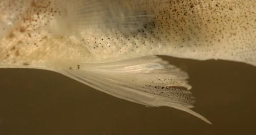 |
| Lutjanus
cyanopterus juvenile |
| 30.2 mm SL, DNA-confirmed
ID |
| dark variation |
| note enlarged canines |
| Isla Grande, Panama,
N7527b |
|
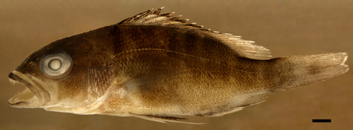 |
| |
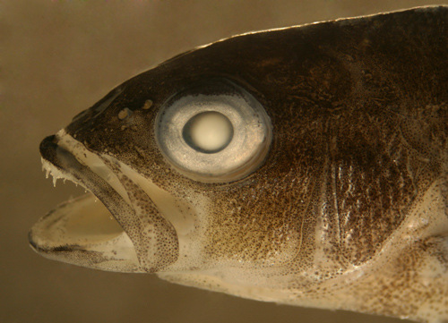 |
| Lutjanus
cyanopterus juvenile |
| 34.0 mm SL, DNA-confirmed
ID |
| light variation |
| very indistinct bars |
| Isla Grande, Panama,
N7529a |
|
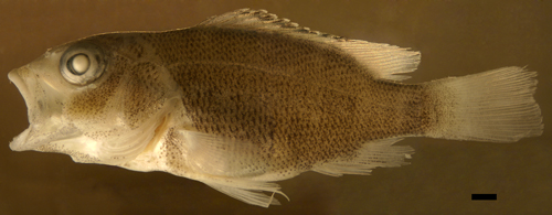 |
| |
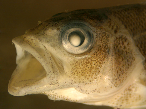 |
| Lutjanus
cyanopterus spawning adult |
| about one meter TL,
returned alive |
| St. Thomas, USVI, 2006 |
|
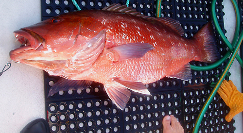 |
|
|
|
|
| |
|
|
|
| Diagnosis:
Modal fin-ray counts of D-X,14 A-III,8 are
shared among most of the regional Lutjanus species,
including L.
analis, L.
apodus, L.
cyanopterus, L.
griseus, L.
jocu and the deep-water snappers L. buccanella,
L.
campechanus, and L.
vivanus. L. buccanella juveniles
have a distinctive yellow saddle mark on the upper
caudal peduncle and an indistinct lateral spot.
(DNA) |
|
| Description:
Body wide and relatively thick with a sloping
forehead and a large round eye (distinctly larger
than in the shallow-water congeners) and large terminal
mouth. Dorsal-fin base long and anal-fin base short.
Prominent dorsal, anal, and pelvic-fin spines and
a large non-serrated preopercular spine. |
|
Transitional
stage:
Transitional recruits show only a speckling of fine
surface melanophores in addition to the residual
larval melanophore complement. The lateral spot
is made up of fine melanophores in a wide ellipse
centered on the lateral line under the soft dorsal
fin. The body becomes covered in fine leukophores.
Lateral and ventral iridescence is prominent and
includes a stripe from the mid-upper jaw around
the lower eye socket and then widening to cover
the preopercle. In life, a distinctive yellow saddle-patch
develops on the dorsal caudal peduncle. |
|
Juveniles:
Juvenile L. buccanella |
|
|
|
| Lutjanus
buccanella transitional recruit |
| 24.5 mm SL, DNA-confirmed
ID |
| note indistinct lateral
spot at lateral line |
| San Blas, Panama, SB82-005 |
|
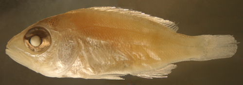 |
| |
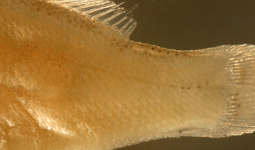 |
| |
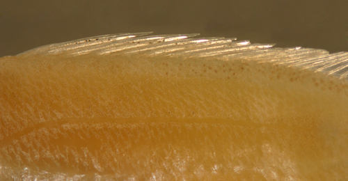 |
|
|
|
|
| |
|
|
|
|
|
|
| |
|
|
|
|
| |
 |
|
All contents © copyright 2006-2015
All rights reserved
www.coralreeffish.com by
Benjamin Victor
|
|
|
|
|
|