The
snake and worm eels comprise the largest eel
family with at least 44 species in the broadly
defined Caribbean region. Despite the large
number of species, these eels are rarely seen
underwater and are found mainly on deeper
soft sediments where some are frequently collected
in shrimp trawls. Many species, however, are
apparently rare and known from few individuals.
Nevertheless, their leptocephalus larvae are
often not rare, indicating that collection
methods for adult ophichthids are generally
suboptimal and there is a lot more to be discovered.
The diversity of leptocephali in the region
is clearly greater than the known set of adults
and resolving the larvae should assist in
completing the taxonomy for the group.
The
state of the art of the taxonomy and larval
identification of eels was captured in 1989
by the book of the Fishes of the Western Atlantic,
Part 9. This comprehensive review of adult
taxonomy, with the ophichthids covered by
John McCosker and Eugenia and James Bohlke,
reviewed all the known species in the region.
The companion volume on the leptocephali,
with ophichthids covered by Mark Leiby, described
the larvae of most of the known species and
included many larval types unassigned to species.
At the time, the primary method to identify
leptocephali was to match meristics- a difficult
task given the number of similar species and
the undiscovered species within several groups.
The advent of DNA identification of larvae
heralded a new age in larval identifications;
however, molecular IDs required new material
to be collected, since formalin preservation
precludes DNA sequencing. The necessity for
new collections has retarded progress on the
identification of leptocephali, but now a
large set of leptocephali from the Yucatan
region have been recently collected in ethanol
and barcoded by the ECOSUR project led by
Lourdes Vasquez-Yeomans and Martha Valdez
and another large set from the Florida Straits
collected by David Richardson of the Northeast
Fisheries Science Center/ NMFS/NOAA is now
being barcoded. In addition, my smaller collections
of leptocephali from Belize and Panama, as
well as my comparative collections of the
sibling species from the eastern Pacific are
also being DNA barcoded. Without question,
the key to resolving the taxonomy of these
eels will be genetic material from adult specimens
for comparison, although larval sequences
alone can reveal near-relatives and thus assign
types to genera, at least. These collections
are likely to resolve most of the questions
raised in the 1989 review and provide a remarkably
comprehensive description of the ophichthid
larvae from the New World.
The
descriptions and keys in the Leiby chapter
in the FWNA book emphasize meristics and morphometrics,
since those were the more useful characters
to connect larval stages with adult stages
at the time. Melanophore patterns were less
emphasized and the line drawings frequently
obscured or did not illustrate melanophores,
especially on the head and caudal patterns.
I emphasize marking patterns more in this
page, since my objective is to facilitate
visual recognition rather than connecting
larvae with adults, which is easier done by
DNA matching these days. In general, for mature
leptocephali, species can be distinguished
by unique combinations of melanophore patterns
and the more tedious and difficult myomere
counts are presented simply to confirm the
species identification provided by DNA matching.
I use some different terminology to describe
melanophore patterns to make them more specific
and easier to abbreviate (described in detail
below). In addition, my treatment differs
from the pigmentation sections in the Leiby
chapter in that I prefer to describe the complement
of melanophores on mature leptocephali and
present a separate account for immature larvae
of each species. The format in the FWNA book
presents markings on each part of the body
separately for early, immature, and mature
stages (as early engyodontic through late
eurydontic stages) before going on to the
next part of the body- making it difficult
to tease out the overall marking pattern for
mature larvae from the text.
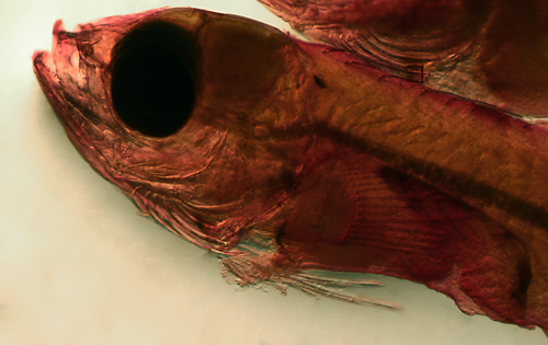
Melanophore
patterns in Ophichthidae
Ophichthid leptocephali share some
basic melanophore patterns that are very useful for identification.
Most obviously, they have multiple swellings along the
gut, usually capped with melanophores, and the number
of these swellings is an important character. A variety
of species-specific melanophore patterns are centered
around these swellings. Almost all ophichthid leptocephali
also have a set of internal melanophore patches along
the tail (defined as postanal), below the notochord (the
rod of developing vertebrae, below the nerve cord which
is orange in the photo at right), called sub-notochordal
caps (SNC), and the number of these are similarly important
for identification. Why develop melanophore coverings
along the tail below the developing nerve cord and notochord?
This location on zebrafish embryos comprises the caudal
hematopoetic tissue (CHT): perhaps these vulnerable cells
are concentrated in patches and need to be shielded from
UV rays (right, lower). In addition to these basic marking
patterns, a variety of additional patterns of melanophores
on the head, thoracic organs, lateral midline, and dorsal
and ventral margins are critical distinguishing features.
Each species tends to have a unique combination of these
patterns. The number and placement of these melanophores
will identify ophichthine leptocephali to species most
of the time. The types of melanophore patterns are listed
below in rough order of decreasing significance.
Swelling
caps, sides, and ventrum: All ophichthid leptocephali
have gut swellings to some degree, although the intestinal
swellings after the liver swellings can sometimes be much
reduced. Nevertheless, the number of swellings and the
markings on them are important identification features.
The first two or three swellings comprise liver and gallbladder
and have a different form than the subsequent gut swellings
(detailed in the description of myrophins and ophichthins
below). A swelling can have a variety of associated melanophore
patterns. At the top there can be an internal dorsal nephric
cap of melanophores, covering the nephric tube above the
intestine. A common marking is a cap-like layer of melanophores
over the intestine itself, below the nephric tube, which
frequently extends laterally and appears to be a line
under the nephric cap. Less frequently there can be some
independent surface melanophores on the side of the swelling
(not part of the dorsal cap) or on the ventral aspect.
(NSC=nephric swelling cap, GSC=gut swelling cap, SS=swelling
side, SV=swelling ventral)
Gut
loop melanophores: Most species have hanging loops
of gut between swellings, although some species have very
shallow or almost indistinct loops. Melanophores on these
loops can be placed over the gut, below the nephros, or
on the ventral surface. The pattern is usually one midway
between gut swellings (rarely two, sometines an irregular
line). These melanophores need to be distinguished from
nephric or gut swelling caps when the loops are low and
indistinct or they will result in overcounting of the
number of gut swellings. Some species have these only
on the first, or esophageal, gut loop. (DGL=dorsal gut
loop, VGL=ventral gut loop, VEGL=ventral esophageal gut
loop)
Sub-notochordal
cap: These deep internal melanophores occur on
almost all ophichthid species (with the notable exception
of Myrophis
punctatus and M. plumbeus from south of
the Caribbean). They appear as short lines slightly below
the lateral midline, sloping down and rearward or running
horizontal, and each spanning about two myomeres. Typically
the patches are spaced well apart along the tail (i.e.
postanal; although in Myrophis
platyrhynchus and some Pseudomyrophis,
the first is characteristically just preanal). They are
arranged on the membrane covering the central tissue mass
below the notochord and form a plane, sometimes with long
filamentous dorsal extensions. In the FWNA these melanophores
are referred to as "subcutaneous pigment patches
on tail just ventral to notochord". They are frequently
associated with surface melanophores, either streaks along
the myoseptum and/or patches. (SNC=sub-notochordal cap)
Spinal
cap: In a few species (especially Callechelynae),
there are deep patches of melanophores draped over the
nerve cord and notochord, slightly above the midline.
These patches are usually limited to the body (i.e. preanal),
span about two myomeres each, and are spaced well apart.
They are frequently associated with surface melanophores,
either in streaks, patches, or both. (SPC=spinal cap)
Myoseptal
streaks: The number and form of surface melanophores
along the lateral midline are important characters. Most
common are myoseptal streaks: thin lines of small melanophores,
usually merged, along the crease between myomeres (the
myoseptum). These streaks can be short or long, single
or multiple. They can be on every myoseptum or spaced
out to some degree, with the number of unmarked myosepta
between streaks (or pairs or triplets of streaked myosepta),
as well as the overall percentage of streaked myosepta,
very useful characters. Frequent patterns are short-long-short
(over each SNC) with another single short spaced about
5 myomeres away (sls5s5sls). Short is about the length
of one myomere-width, long is about two or three. The
streak series are typically centered slightly below the
lateral midline. (MS=myoseptal streak, s=short, l=long)
Lateral
midline patches: While streaks are most common,
there can be patches of small melanophores either in place
of, or in addition to, the myoseptal streaks along the
surface at the lateral midline (or, rarely, single larger
melanophores). The patches can be irregular, sometimes
just a few extra melanophores around a streak, or in various
degrees of organization up to discrete round collections.
Rarely there are patches away from the midline, either
above the gut loops or above the anal fin (LMP=lateral
midline patch, LM1=lateral midline single spot)
Anal
fin base: The anal fin typically starts right after
the anus and runs along the ventral midline. Two distinct
patterns of small melanophores can line the base of the
pterygiophores, usually one per element: either a continuous
line or a broken series of short rows of melanophores
broken by unmarked gaps. Many species' leptocephali have
no anal fin melanophores, although a few have them only
near the very end of the tail (in this discussion considered
caudal patterns). A rare pattern is saddle-like patches
of melanophores spaced out widely along the base. (AFB=anal
fin base, cont or broken or saddles)
Dorsal
midline: Most leptocephali have no dorsal midline
melanophores, but some species have a row of tiny melanophores,
often one at each dorsal-fin pterygiophore and, if continuing
forward of the fin origin, usually spaced farther apart.
A rare pattern is a series of rounded patches of small
melanophores spaced out widely along the dorsal midline.
As with the anal fin, some species without dorsal midline
arrays will have a few small melanophores at the base
of the last few dorsal fin elements at the tail (in this
discussion considered caudal patterns). (DM=dorsal midline
series, row or patches)
Pharyngeal
and cardiac: Thoracic patterns typically comprise
a cap of deep melanophores over the pharynx (just forward
of the pectoral fins) and/or lateral or ventral cardiac
melanophores. The pharyngeal cap technically is also covering
the beginning of the esophagus, but I reserve the label
esophagus for the first gut loop. There can be a patch
ventral to the pharyngeal/esophageal area over the cardiac
bulge. Cardiac melanophores include internal aortic coverings
(forward of the bulge) or surface melanophores at the
side (often in a linear series) or in a patch on the ventral
surface. (PC=pharyngeal cap; VP=ventral pharyngeal, AO=aortic,
CS=cardiac side or CV=cardiac ventral)
Cranial melanophores:
A variety of patterns of melanophores are found on the
head, sometimes including deep melanophores lining the
lower cranial vault around the brain, perhaps protecting
the haircells of the semicircular canals. (B=braincase)
Maxillary
and mandibular: Deep small melanophores are frequently
located along the maxilla on each side, below the base
of the teeth, and concentrated along the anterior third
of the maxilla: they are frequently in a row or just one
or two. Lower-jaw melanophores are usually concentrated
near the tip of the jaw. (MAX= maxillary row, LJ=tip of
lower jaw)
Caudal
melanophores: The pattern of melanophores at the
end of the tail does not vary much between species and
typically comprises a short line over the end of the spinal
cord, a short line or row of discrete small melanophores
under the notocord, and frequently a few melanophores
at the base of the very last dorsal and anal fin elements.
In some species with well spaced-out lateral midline streaks,
the last few myomeres at the tail can have streaks on
each myoseptum. Infrequently melanophores are present
on the caudal fin membranes (CSC=over caudal spinal cord,
CSN=caudal sub-notochord, CP=caudal pterygiophore bases,
CM=caudal fin membranes)
Subfamily
Myrophinae
This relatively small subset of the
family comprise the worm eels and have larvae that are
easily distinguished by having a unique arrangement of
gut swellings, i.e. three liver swellings with the gall
bladder on the third (vs. only two in other ophichthids).
Notably the gut coming into the third swelling is thin
and becomes distinctly wider as it leaves. Adults are
less distinctive and often hard to quickly identify as
myrophins, although they do have a visible caudal fin
with fin rays, while other snake and worm eels often have
a finless point to the tail.
There are four genera of tropical
Western Atlantic myrophins with about eight species
in the region at present. However, based on available
leptocephali there may be more species and certainly
the known ranges are uncertain. This group of eels
are particularly difficult to sample adequately,
perhaps because they are smaller than most net meshes,
live in deep and rarely accessed habitats and may
be buried in sand most of the time. Ahlia
egmontis and Myrophis
punctatus are common and widespread in shallow
Caribbean waters, although they are rarely seen
by divers.
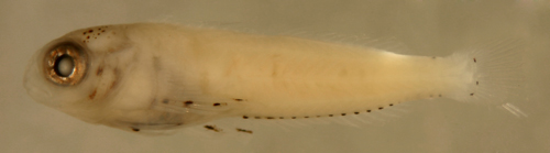
Ahlia egmontis
Identification:
Three liver swellings plus 1-5 more gut swellings,
indistinct gut loops, dorsal midline melanophores,
short streaks on almost every myoseptum.
Meristics: MYO:
total 152-168, nephric 64-73, preanal 67-75, predorsal
65-76. DFO: 70. Nephros ends -3PA. Reach 90-100
mm.
Description:
Gut with . Body with DMrow, MS q1 after myo5
(short), 5 SNC, AFBcont. Head with . Caudal with
.
Ahlia
egmontis
18.9 mm SL
San Blas, Panama, SB86-422
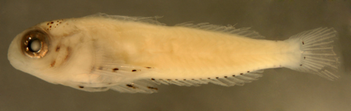
Myrophis punctatus
Identification:
Three liver swellings followed by mostly straight
gut. Single melanophores, occasionally two, along
lateral midline on a third to half of the myosepta.
No sub-notochordal caps, no dorsal midline melanophores.
Meristics: MYO:
total 137-152, nephric 53-60, preanal 53-62, predorsal
30-38. DFO: 30. Nephros ends -1PA. Reach 80-90
mm.
Description:
Gut with . Body with LM1 q 1-3, no SNC, AFBcont.
Head with . Caudal with .
Hypsoblennius
invemar larva
11.4 mm SL
San Blas, Panama, SB86-422
Myrophis platyrhynchus
Identification:
Three liver swellings plus 2 or 3 more gut swellings
(5 or 6 total), low gut loops, first SNC before
anus, short myoseptal streaks q 6-8. No dorsal
midline melanophores.